. 2018 Jan;32(1):488-499.
doi: 10.1096/fj.201700670R. Epub 2017 Sep 22.

α-Ketoglutarate prevents skeletal muscle protein degradation and muscle atrophy through PHD3/ADRB2 pathway

Affiliations

α-Ketoglutarate prevents skeletal muscle protein degradation and muscle atrophy through PHD3/ADRB2 pathway

Xingcai Cai et al. FASEB J. 2018 Jan.

Erratum in

Abstract

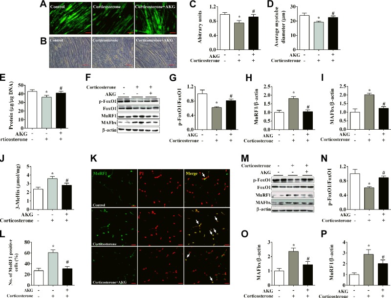
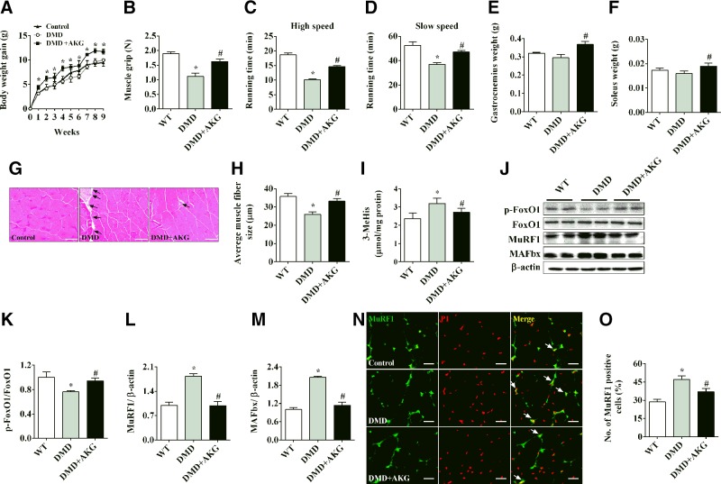
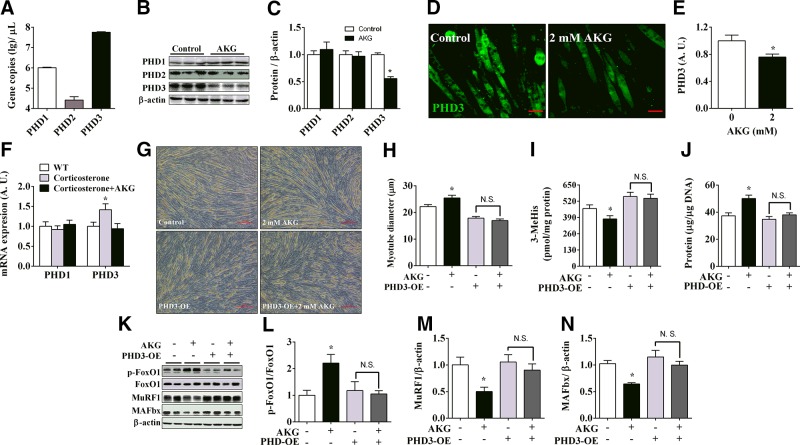
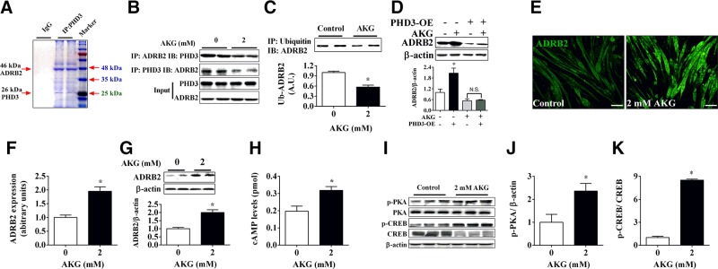
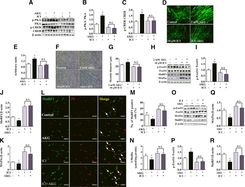
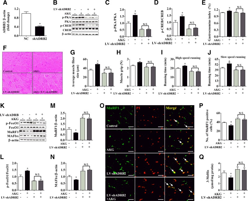
Skeletal muscle atrophy due to excessive protein degradation is the main cause for muscle dysfunction, fatigue, and weakening of athletic ability. Endurance exercise is effective to attenuate muscle atrophy, but the underlying mechanism has not been fully investigated. α-Ketoglutarate (AKG) is a key intermediate of tricarboxylic acid cycle, which is generated during endurance exercise. Here, we demonstrated that AKG effectively attenuated corticosterone-induced protein degradation and rescued the muscle atrophy and dysfunction in a Duchenne muscular dystrophy mouse model. Interestingly, AKG also inhibited the expression of proline hydroxylase 3 (PHD3), one of the important oxidoreductases expressed under hypoxic conditions. Subsequently, we identified the β2 adrenergic receptor (ADRB2) as a downstream target for PHD3. We found AKG inhibited PHD3/ADRB2 interaction and therefore increased the stability of ADRB2. In addition, combining pharmacologic and genetic approaches, we showed that AKG rescues skeletal muscle atrophy and protein degradation through a PHD3/ADRB2 mediated mechanism. Taken together, these data reveal a mechanism for inhibitory effects of AKG on muscle atrophy and protein degradation. These findings not only provide a molecular basis for the potential use of exercise-generated metabolite AKG in muscle atrophy treatment, but also identify PHD3 as a potential target for the development of therapies for muscle wasting.-Cai, X., Yuan, Y., Liao, Z., Xing, K., Zhu, C., Xu, Y., Yu, L., Wang, L., Wang, S., Zhu, X., Gao, P., Zhang, Y., Jiang, Q., Xu, P., Shu, G. α-Ketoglutarate prevents skeletal muscle protein degradation and muscle atrophy through PHD3/ADRB2 pathway.

Keywords: Duchenne muscular dystrophy; metabolism; metabolite; tricarboxylic acid cycle.


Conflict of interest statement

This work was supported by the National Basic Research Program of China (Grant 2013CB127306 to G.S.), National Key Point Research and Invention Program (Grant 2016YFD0501205 to G.S.), Training Program for Outstanding Young Teachers in the Universities of Guangdong Province (G.S.), National Natural Science Foundation of China (Grants 31572480 to G.S. and 31472105 to Q.J.), and the U.S. National Institutes of Health, National Institute of Diabetes and Digestive and Kidney Diseases (Grant K99DK107008 to P.X.). The authors declare no conflicts of interest.

Figures

Figure 1.
Figure 1.
AKG inhibits corticosterone-induced protein degradation in skeletal muscle. A, C) Representative images (A) and fluorescence intensity quantification (C) of long-lived protein Click-It AHA in C2C12 myotubes treated with vehicle, 10 μM corticosterone, or 10 μM corticosterone + 2 mM AKG for 48 h. B, D) Representative images (B) and quantification (D) of C2C12 myotubes size treated for 48 h. E) Total protein levels of C2C12 myotubes treated for 48 h. FI) Immunoblots (F) and quantification (GI) of pFoxO1 and FoxO1 (G), MuRF1 (H), and MAFbx (I) in C2C12 myotubes treated for 48 h. J) 3-MeHis levels in gastrocnemius muscle from male C57BL/6J mice injected intraperitoneally with vehicle, 50 μg/kg corticosterone, or 50 μg/kg corticosterone + 1.0 g/kg AKG. 3-MeHis levels were detected by HPLC to measure protein degradation. K, L) Representative images (K) and quantification (L) of propidium iodide (PI, red)–positive MuRF1 (green) cells (yellow nucleus indicated by white arrows) in gastrocnemius muscle from male C57BL/6J mice injected intraperitoneally with vehicle, corticosterone, or corticosterone + AKG. MP) Immunoblots (M) and quantification (NP) of pFoxO1 and FoxO1 (N), MuRF1 (O), and MAFbx (P) in gastrocnemius muscle from male C57BL/6J mice injected intraperitoneally with vehicle, corticosterone, or corticosterone + AKG. All results contain 6 replicates per group (n = 6/group). Data are presented as means ± sem and were analyzed by 1-way ANOVA, followed by post hoc Bonferroni tests. β-Actin served as housekeeping gene. *P < 0.05 compared to control, #P < 0.05 compared to corticosterone group.
Figure 2.
Figure 2.
AKG rescues skeletal muscle atrophy in mdx mice. A) Body weight of male C57BL/6J mice receiving tap water, mdx mice receiving water, or mdx mice receiving water supplemented with 2% AKG for 8 wk. B) Muscle grip of male mice provided water or 2% AKG for 8 wk. C, D) Treadmill running time of male mice provided water or 2% AKG for 8 wk at high-speed (C) and slow-speed (D) settings. E, F) Gastrocnemius (E) and soleus (F) muscle weight of male mice provided water or 2% AKG for 8 wk. G, H) Representative images (G) and quantification (H) of gastrocnemius muscle hematoxylin and eosin staining from male mice provided water or 2% AKG for 8 wk. I) Levels of 3-MeHis in gastrocnemius muscle from male mice provided water or 2% AKG for 8 wk. JM) Immunoblots (J) and quantification (KM) of pFoxO1 and FoxO1 (K), MuRF1 (L), and MAFbx (M) in gastrocnemius muscle from male mice provided water or 2% AKG for 8 wk. N, O) Representative images (N) and quantification (O) of propidium iodide (PI, red)-positive MuRF1 (green) cells (yellow nucleus indicated by white arrows) in gastrocnemius muscle from male mice provided water or 2% AKG for 8 wk. All results contain 10 replicates per group (n = 10/group). Data are presented as means ± sem and were analyzed by 1-way ANOVA, followed by post hoc Bonferroni tests. β-Actin served as housekeeping gene. *P < 0.05 compared to control; #P < 0.05 compared to MDX group.
Figure 3.
Figure 3.
PHD3 mediates inhibitory effects of AKG on protein degradation and myotube atrophy. A) C2C12 cells were induced for 4 d to differentiate into myotubes. Then C2C12 myotubes were collected for examining expression of 3 PHDs subtypes by absolute quantification PCR (n = 6/group). B, C) Immunoblots (B) and quantification (C) of PHDs in C2C12 myotubes treated with 0 or 2 mM AKG for 48 h (n = 6/group). D, E) Representative images (D) and quantification (E) of PHD3 in C2C12 myotubes treated with 0 or 2 mM AKG for 48 h (n = 6/group). F) mRNA expression of PHD1 and 3 in gastrocnemius muscle from 8 wk old male C57BL/6J mice injected intraperitoneally with vehicle, 50 μg/kg corticosterone, or 50 μg/kg corticosterone + 1.0 g/kg AKG (n = 6/group). G, H) Representative images (G) and quantification (H) of diameters of normal or PHD3-overexpressing C2C12 myotubes treated with 2 mM AKG for 48 h (n = 6/group). I) 3-MeHis levels in culture medium from normal or PHD3-overexpressing C2C12 myotubes treated with 2 mM AKG for 48 h (n = 6/group). J) Total protein levels in normal or PHD3-overexpressing C2C12 myotubes treated with 2 mM AKG for 48 h (n = 6/group). KN) Immunoblots (K) and quantification (LN) of pFoxO1 and FoxO1 (L), MuRF1 (M), and MAFbx (N) in normal or PHD3-overexpressing C2C12 myotubes treated with 2 mM AKG for 48 h (n = 6/group). Data are presented as means ± sem and were analyzed by 1-way ANOVA, followed by post hoc Bonferroni tests. β-Actin served as housekeeping gene. *P < 0.05 compared to control.
Figure 4.
Figure 4.
ADRB2 is target of PHD3 in response to AKG. A) C2C12 cells were induced differentiation for 4 d, and anti-PHD3 was used to pull down PHD3 binding protein. Precipitated samples were used for SDS-PAGE, and then strips were stained by Coomassie Brilliant Blue. B) C2C12 myotubes were treated by 2 mM AKG for 48 h, and anti-PHD3 and anti-ADRB2 were used to precipitate PHD3 and ADRB2, respectively. Precipitated samples was subjected to immunoblotting of ADRB2 or PHD3 (n = 3 group). C) C2C12 myotube samples from control or 2 mM AKG–treated group were pulled down by anti-ubiquitin antibody and subjected to immunoblotting of ADRB2 (n = 3/group). D) Immunoblots and quantification of ADRB2 in normal or PHD3-overexpressing C2C12 myotubes treated with 2 mM AKG for 48 h (n = 6/group). E, F) Representative images (E) and quantification (F) of ADRB2 in C2C12 myotubes treated with 0 or 2 mM AKG for 48 h (n = 6/group). G) Immunoblots and quantification of ADRB2 in C2C12 myotubes treated with 0 or 2 mM AKG for 48 h (n = 6/group). H) cAMP levels in C2C12 myotubes treated with 0 or 2 mM AKG for 48 h (n = 6/group). IK) Immunoblots (I) and quantification (J, K) of p-PKA (J), PKA, p-CREB (K), and CREB (K) in C2C12 myotubes treated with 0 or 2 mM AKG for 48 h (n = 6/group). Data are presented as means ± sem and were analyzed by 1-way ANOVA followed by post hoc Bonferroni tests for panel D, and nonpaired Student’s t test for all others. β-Actin served as housekeeping gene. *P < 0.05 compared to control.
Figure 5.
Figure 5.
Pharmacologic inhibition of ADRB2 blocked inhibitory effects of AKG on skeletal muscle atrophy and protein degradation. AC) Immunoblots (A) and quantification (B, C) of p-PKA and PKA (B) and p-CREB and CREB (C) in C2C12 myotubes treated with vehicle, 2 mM AKG, 10 μM ICI, or 2 mM AKG + 10 μM ICI for 48 h (n = 6/group). D, E) Representative images (D) and quantification (E) of long-life protein Click-It AHA in C2C12 myotubes treated with vehicle, 2 mM AKG, 10 μM ICI, or 2 mM AKG + 10 μM ICI for 48 h (n = 6/group). F, G) Representative images (F) and quantification (G) of fiber diameter of C2C12 myotubes treated with vehicle, 2 mM AKG, 10 μM ICI, or 2 mM AKG + 10 μM ICI for 48 h (n = 6/group). HK) Immunoblots (H) and quantification (IK) of pFoxO1 and FoxO1 (I), MuRF1 (J), and MAFbx (K) in C2C12 myotubes treated with vehicle, 2 mM AKG, 10 μM ICI, or 2 mM AKG + 10 μM ICI for 48 h (n = 6/group). L, M) Representative images (L) and quantification (M) of propidium iodide (PI, red)-positive MuRF1 (green) cells (yellow nucleus indicated by white arrows) in gastrocnemius muscle from male C57BL/6J mice 3 h after intraperitoneal injection with vehicle, 1 g/kg AKG, 5 μg/kg ICI, or 1 g/kg AKG + 5 μg/kg ICI (n = 6). N) 3-MeHis in gastrocnemius muscle from male C57BL/6J mice 3 h after intraperitoneal injection with vehicle, 1 g/kg AKG, 5 μg/kg ICI, or 1 g/kg AKG + 5 μg/kg ICI (n = 6). OR) Immunoblots (O) and quantification (PR) of pFoxO1 and FoxO1 (P), MuRF1 (Q), and MAFbx (R) in gastrocnemius muscle from male C57BL/6J mice 3 h after injected intraperitoneally with vehicle, 1 g/kg AKG, 5 μg/kg ICI, or 1 g/kg AKG + 5 μg/kg ICI (n = 6). Data are presented as means ± sem and were analyzed by 1-way ANOVA, followed by post hoc Bonferroni tests. β-Actin served as housekeeping gene. *P < 0.05 compared to control.
Figure 6.
Figure 6.
ADRB2 knockdown abolished inhibitory effects of AKG on muscle atrophy and protein degradation. A) mRNA expression of ADRB2 in gastrocnemius muscle from male C57BL/6J mice injected intramuscularly with LV-shScrambled or LV-shADRB2. BD) Immunoblots (B) and quantification (C, D) of p-PKA and PKA (C) and p-CREB and CREB (D) in gastrocnemius muscle from male C57BL/6J mice receiving LV-shScrambled, LV-shScrambled + AKG, LV-shADRB2, or LV-shADRB2 +AKG. EJ) Gastrocnemius weight (E), gastrocnemius muscle fiber size (F, G), muscle grip (H), high-speed running time (I), and slow-speed running time (J) of male C57BL/6J mice receiving LV-shScrambled, LV-shScrambled + AKG, LV-shADRB2, or LV-shADRB2 + AKG. KN) Immunoblots (K) and quantification (LN) of pFoxO1 and FoxO1 (L), MuRF1 (M), and MAFbx (N) in gastrocnemius muscle from male C57BL/6J mice receiving LV-shScrambled, LV-shScrambled + AKG, LV-shADRB2, or LV-shADRB2 + AKG. O, P) Representative images (O) and quantification (P) of propidium iodide (PI, red) positive MuRF1 (green) cells (yellow nucleus indicated by white arrows) in gastrocnemius muscle from male C57BL/6J mice receiving LV-shScrambled, LV-shScrambled + AKG, LV-shADRB2, or LV-shADRB2 + AKG. Q) Levels of 3-MeHis in gastrocnemius muscle from male C57BL/6J mice receiving LV-shScrambled, LV-shScrambled + AKG, LV-shADRB2, or LV-shADRB2 + AKG. Data are presented as means ± sem and were analyzed by nonpaired Student’s t test for panel A, and 1-way ANOVA, followed by post hoc Bonferroni tests for all others. β-Actin served as housekeeping gene. *P < 0.05 compared to control.

References

    1. Bonaldo P., Sandri M. (2013) Cellular and molecular mechanisms of muscle atrophy. Dis. Model. Mech. 6, 25–39 - PMC - PubMed
    1. Malicdan M. C., Noguchi S., Nonaka I., Saftig P., Nishino I. (2008) Lysosomal myopathies: an excessive build-up in autophagosomes is too much to handle. Neuromuscul. Disord. 18, 521–529 - PubMed
    1. Stephens N. A., Gallagher I. J., Rooyackers O., Skipworth R. J., Tan B. H., Marstrand T., Ross J. A., Guttridge D. C., Lundell L., Fearon K. C., Timmons J. A. (2010) Using transcriptomics to identify and validate novel biomarkers of human skeletal muscle cancer cachexia. Genome Med. 2, 1. - PMC - PubMed
    1. Gordon B. S., Kelleher A. R., Kimball S. R. (2013) Regulation of muscle protein synthesis and the effects of catabolic states. Int. J. Biochem. Cell Biol. 45, 2147–2157 - PMC - PubMed
    1. Verdijk L. B., Dirks M. L., Snijders T., Prompers J. J., Beelen M., Jonkers R. A., Thijssen D. H., Hopman M. T., Van Loon L. J. (2012) Reduced satellite cell numbers with spinal cord injury and aging in humans. Med. Sci. Sports Exerc. 44, 2322–2330 - PubMed

Publication types

MeSH terms