. 2017 Nov 6;216(11):3453-3462.
doi: 10.1083/jcb.201705168. Epub 2017 Sep 22.

Regulation of mitotic spindle assembly factor NuMA by Importin-β

Affiliations

Regulation of mitotic spindle assembly factor NuMA by Importin-β

Chih-Chia Chang et al. J Cell Biol. .

Abstract

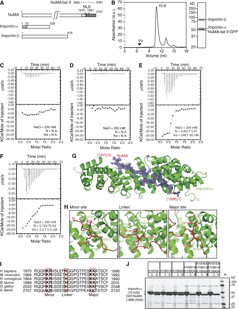
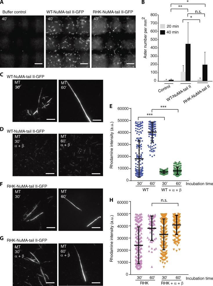
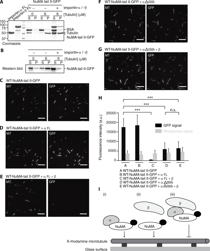
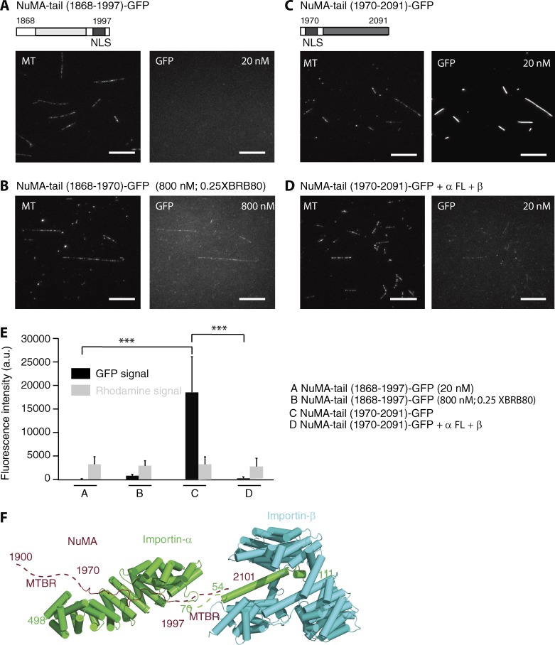
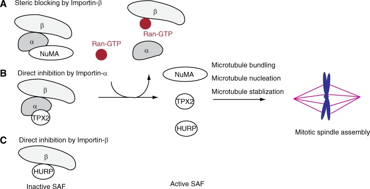
Ran-guanosine triphosphatase orchestrates mitotic spindle assembly by modulation of the interaction between Importin-α/-β and spindle assembly factors (SAFs). The inhibition of SAFs performed by importins needs to be done without much sequestration from abundant nuclear localization signal (NLS) -containing proteins. However, the molecular mechanisms that determine NLS-binding selectivity and that inhibit activity of Importin-β-regulated SAFs (e.g., nuclear mitotic apparatus protein [NuMA]) remain undefined. Here, we present a crystal structure of the Importin-α-NuMA C terminus complex showing a novel binding pattern that accounts for selective NLS recognition. We demonstrate that, in the presence of Importin-α, Importin-β inhibits the microtubule-binding function of NuMA. Further, we have identified a high-affinity microtubule-binding region that lies carboxyl-terminal to the NLS, which is sterically masked by Importin-β on being bound by Importin-α. Our study provides mechanistic evidence of how Importin-α/-β regulates the NuMA functioning required for assembly of higher-order microtubule structures, further illuminating how Ran-governed transport factors regulate diverse SAFs and accommodate various cell demands.


Figures

Figure 1.
Figure 1.
Biochemical and structural characterization of the heterotrimeric NuMA-tail IIImportin-αImportin-β complex. (A) Domain structures of NuMA, Importin-α, and Importin-β. For NuMA, the microtubule-binding regions suggested by the Merdes and Mapelli groups are shown in light and dark gray, respectively (Haren and Merdes, 2002; Gallini et al., 2016). The NLS is colored black. Residue numbers of NuMA-tail II (dotted line) used in biochemical studies are indicated. For Importin-α, the Importin-β binding domain is indicated. (B) Purified recombinant Importin-α, Importin-β, and NuMA-tail II-GFP were mixed and analyzed by SEC. The peak fraction (10.8 ml) was analyzed by SDS-PAGE and stained with Coomassie blue. SEC (Superdex 200) elution profile for the NuMA-tail II-GFP–Importin-α–Importin-β trimeric complex. The void volume (Vo) of the peak fraction and absorbance (a.u.) at 280 nm for the complex is indicated. (C–F) ITC titration curves (top) and binding isotherms (bottom) of full-length Importin-α (C), full-length Importin-β (D), Importin-α (ΔIBB; E), and 1:1 stoichometric mixture of full-length Importin-α and -β with NuMA-tail II-GFP (F). (G) An omit difference (Fo-Fc) map contoured at 2.5 sigma with a superimposed atomic model of the NuMA (1955–2046)–Importin-α (70–498) complex, showing NuMA in red and Importin-α in green. Residues 1,970 to 1,996 in NuMA are assigned. (H) The three panels highlight the interactions at the minor site, linker region, and major site. (I) Sequence alignment of the NuMA-NLS peptide from different species (numbers represent amino acid positions). The conserved minor-site, linker-region and major-site interacting residues are highlighted by red boxes. (J) GST pull-down assays of Importin-α (70–529) with NuMA-tail mutants. GST-fused NuMA-tail (1,868–2,046) mutants were incubated with recombinant Importin-α. Unbound (S) and bound (B) samples were analyzed by SDS-PAGE and stained with Coomassie blue.
Figure 2.
Figure 2.
Importin-α/-β regulates NuMA-tail II-mediated higher-order microtubule structure assembly. (A) Microtubule aster assembly in Xenopus egg extracts, examined with buffer control (left), WT NuMA-tail II-GFP (5 µM, center), and RHK-NuMA-tail II-GFP (5 µM, right) after 40-min incubation at 18°C. Microtubules were imaged using X-rhodamine tubulin (100 nM). Large representative images were created with 3×3 raster scan images. Bars, 100 µm. (B) Dependency of aster assembly activity on NuMA. The number of microtubule asters was counted in microtubule images at indicated incubation times. Data are mean ± SD from n = 6 trials, performed using three independent extract preparations (10 ± 9, 109 ± 76, and 21 ± 14 at 20 min; 12 ± 10, 445 ± 256, and 194 ± 152 at 40 min; for control, WT, and RHK mutant, respectively). (C–H) GMPCPP-stabilized microtubules (X-rhodamine–labeled) were incubated separately with equal concentrations (400 nM) of WT-tail II-GFP (C and D) and RHK-tail II-GFP (F and G) in the presence or absence of Importin-α and Importin-β (4 µM each). Samples were fixed and processed to determine rhodamine fluorescence intensity after 30 and 60 min incubation. Bars, 10 µm. (E and H) Rhodamine intensity of microtubules for each condition was analyzed and plotted. SD was determined from data pooled from three independent experiments (E: WT, 30′, n = 180; WT+α+β, 30′, n = 153; WT, 60′, n = 100; WT+α+β, 60′, n = 147. H: RHK, 30′, n = 180; RHK+α+β, 30′, n = 180; RHK, 60′, n = 55; RHK+α+β, 60′, n = 147). Two-tailed Student t test for comparison of 30 min incubation; Mann-Whitney U test for comparison of 60 min incubation. Statistical differences: *, P < 0.05; **, P < 0.01; ***, P < 0.001; n.s., not significant.
Figure 3.
Figure 3.
Importin-β regulates interaction of NuMA-tail II with microtubules. (A) SDS-PAGE analysis of microtubule cosedimentation assays for NuMA-tail II-GFP in the presence of Importin-α or Importin-α/-β. BSA (final, 0.25 mg/ml), which was used to suppress nonspecific interactions, and tubulin concentrations are indicated. Supernatant and pellet fractions are indicated as S and P, respectively. FL, full length. (B) The NuMA-tail II-GFP bands in the gel shown in A were detected by Western blot using anti-GFP antibody as the positions of NuMA-tail II-GFP and tubulin overlap. (C–G) GMPCPP-stabilized microtubules (X-rhodamine– and biotin-labeled), immobilized on a glass surface, were incubated with WT-NuMA-tail II-GFP (C) in the presence of full-length Importin-α (D), full-length Importin-α and Importin-β (E), Importin-α (ΔIBB; F), and Importin-α (ΔIBB) and full-length Importin-β (G). Bars, 10 µm. (H) Analysis of NuMA (GFP) and microtubule (X-rhodamine) fluorescence signals. Mean fluorescence signals under the different conditions shown in C–G were measured and plotted. SD was determined from data pooled from three independent experiments (n = 200 microtubules for each condition). Two-tailed Student t test; statistical differences: ***, P < 0.001; n.s., not significant. (I) A summary of microtubule binding of NuMA-tail II in the presence of Importin-α/-β. X-rhodamine–labeled microtubules were immobilized on the glass surface and NuMA-tail II-GFP (indicated as NuMA; GFP is shown as a black dot) in the presence of Importin-α and Importin-β was imaged using TIRF microscopy.
Figure 4.
Figure 4.
Importin-β regulates the microtubule-binding region that lies C-terminal to the NuMA NLS. GMPCPP-stabilized microtubules (X-rhodamine– and biotin-labeled) were incubated with NuMA-tail (1868–1997) and NuMA-tail (1970–2091) that contain microtubule-binding regions (light and dark gray) and NLS (black). (A and B) 20 nM and 800 nM of NuMA-tail (1868–1997) —GFP were examined under conditions of 1XBRB80 (A) and 0.25XBRB80 (B), respectively. (C and D) 20 nM of NuMA-tail (1970–2091)—GFP in the absence (C) or presence (D) of Importin-α/-β was analyzed. Bars, 10 µm. (E) Mean fluorescence signals under the different conditions shown in A–D were measured and plotted (n = 200 microtubules for each condition). SD was determined from data pooled from three independent experiments. Two-tailed Student t test; statistical differences: ***, P < 0.001. (F) The microtubule-binding region that lies C-terminal to the NLS (aa 1997–2101; brown dotted line) in NuMA is sterically blocked by Importin-β (light blue) that binds to the IBB domain of Importin-α (green; PDB code: 1QGK). Two microtubule-binding regions (MTBRs) are indicated.
Figure 5.
Figure 5.
Schematic for how Ran–GTP regulates SAF activities required for mitotic spindle assembly. SAFs (e.g., NuMA, TPX2, and HURP) are suppressed by the Ran-governed transport factors Importin-α and -β. Ran–GTP modulates interaction between Importins and SAFs. Free SAFs that regulate distinct microtubule behaviors control organization of the mitotic spindle.

References

    1. Brünger A.T., Adams P.D., Clore G.M., DeLano W.L., Gros P., Grosse-Kunstleve R.W., Jiang J.S., Kuszewski J., Nilges M., Pannu N.S., et al. . 1998. Crystallography & NMR system: A new software suite for macromolecular structure determination. Acta Crystallogr. D Biol. Crystallogr. 54:905–921. 10.1107/S0907444998003254 - DOI - PubMed
    1. Cook A., Bono F., Jinek M., and Conti E.. 2007. Structural biology of nucleocytoplasmic transport. Annu. Rev. Biochem. 76:647–671. 10.1146/annurev.biochem.76.052705.161529 - DOI - PubMed
    1. Desai A., Murray A., Mitchison T.J., and Walczak C.E.. 1999. The use of Xenopus egg extracts to study mitotic spindle assembly and function in vitro. Methods Cell Biol. 61:385–412. 10.1016/S0091-679X(08)61991-3 - DOI - PubMed
    1. Fontes M.R., Teh T., and Kobe B.. 2000. Structural basis of recognition of monopartite and bipartite nuclear localization sequences by mammalian importin-alpha. J. Mol. Biol. 297:1183–1194. 10.1006/jmbi.2000.3642 - DOI - PubMed
    1. Forbes D.J., Travesa A., Nord M.S., and Bernis C.. 2015. Nuclear transport factors: global regulation of mitosis. Curr. Opin. Cell Biol. 35:78–90. 10.1016/j.ceb.2015.04.012 - DOI - PMC - PubMed

MeSH terms