. 2017 Sep 22;7(1):12172.
doi: 10.1038/s41598-017-12491-4.

A Single Sphingomyelin Species Promotes Exosomal Release of Endoglin into the Maternal Circulation in Preeclampsia

Affiliations

A Single Sphingomyelin Species Promotes Exosomal Release of Endoglin into the Maternal Circulation in Preeclampsia

Leonardo Ermini et al. Sci Rep. .

Abstract

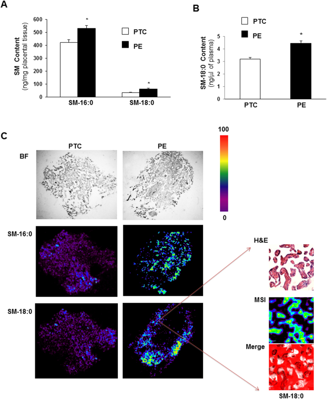
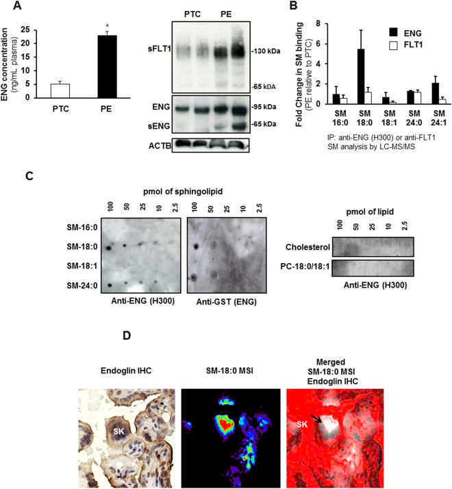
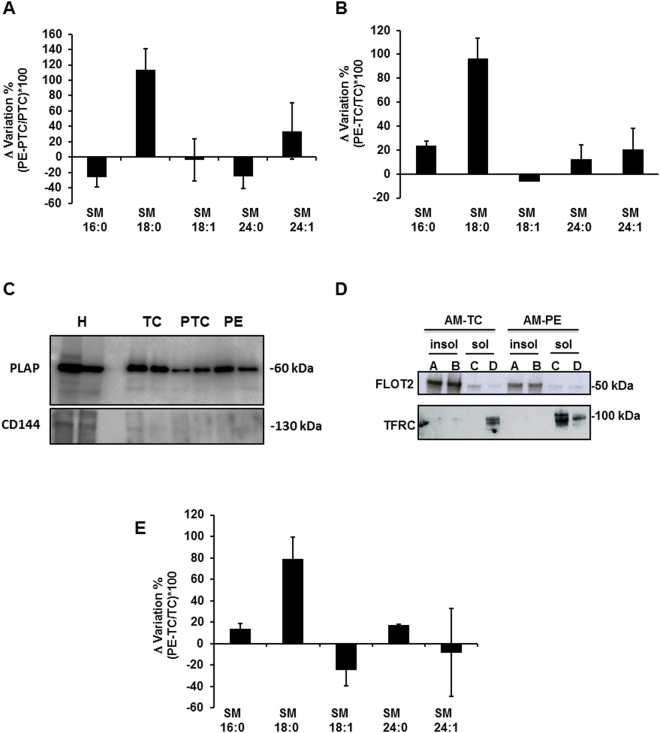
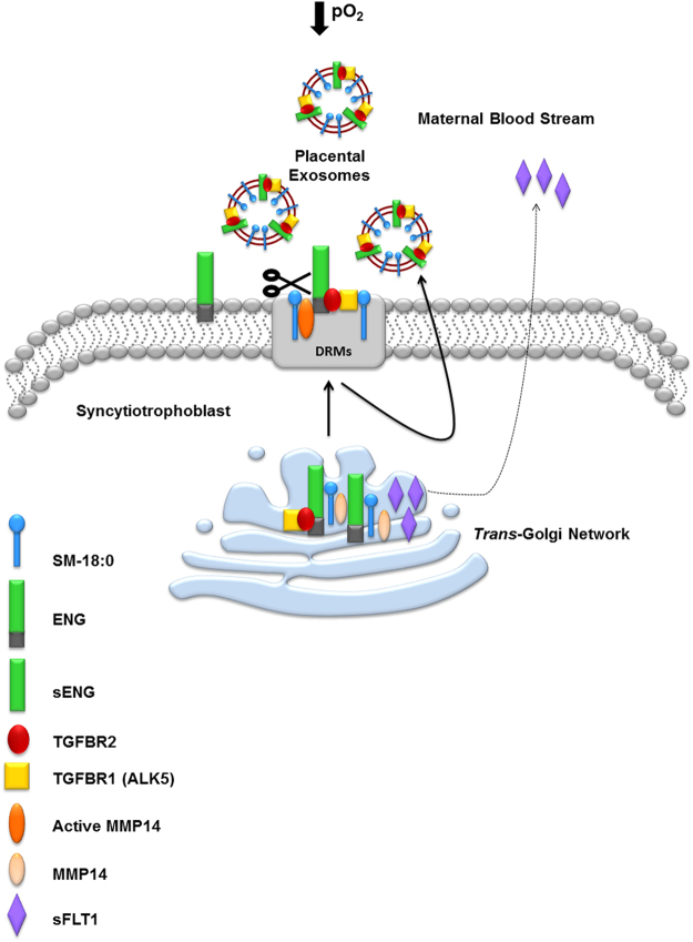
Preeclampsia (PE), an hypertensive disorder of pregnancy, exhibits increased circulating levels of a short form of the auxillary TGF-beta (TGFB) receptor endoglin (sENG). Until now, its release and functionality in PE remains poorly understood. Here we show that ENG selectively interacts with sphingomyelin(SM)-18:0 which promotes its clustering with metalloproteinase 14 (MMP14) in SM-18:0 enriched lipid rafts of the apical syncytial membranes from PE placenta where ENG is cleaved by MMP14 into sENG. The SM-18:0 enriched lipid rafts also contain type 1 and 2 TGFB receptors (TGFBR1 and TGFBR2), but not soluble fms-like tyrosine kinase 1 (sFLT1), another protein secreted in excess in the circulation of women with PE. The truncated ENG is then released into the maternal circulation via SM-18:0 enriched exosomes together with TGFBR1 and 2. Such an exosomal TGFB receptor complex could be functionally active and block the vascular effects of TGFB in the circulation of PE women.


Conflict of interest statement

The authors declare that they have no competing interests.

Figures

Figure 1
Figure 1
Sphingomyelin levels are increased in preeclamptic placentae. (A) Sphingomyelin (SM) levels measured by LC-MS/MS in placental tissue from preeclamptic (PE) women compared to normotensive age-matched preterm controls (PTC). SM numbers indicate fatty acid chain length on D-erythro-sphingosylphosphorylcholine backbone (PE, n = 45; PTC, n = 40 different placentae; *p < 0.05). (B) N-stearoyl sphingomyelin (SM-18:0) levels measured by LC-MS/MS in plasma from PE vs PTC women (PE, n = 10; TC, n = 10 separate samples; *p < 0.05). Data are presented as mean ± s.e.m. Significance was determined using an unpaired two-sided t-test. (C-left panels) Representative MALDI-MS images of SM-16:0 and SM-18:0 distributions in PE and PTC placental sections. Intensities of the ions based on the intensity scale provided. All imaging experiments were repeated with tissues obtained from 6 different PE and PTC placentae. (C-right panels) H&E and MALDI-MS images of SM-18:0 in PE placental terminal villi (magnification: 40x). White pseudocolor indicates overlap between MSI SM-18:0 and H&E stained terminal villi. Red is chosen as pseudocolor background.
Figure 2
Figure 2
Endoglin associates with SM-18:0 in preeclamptic placenta. (A-left panel) ENG content (mean ± s.e.m.) measured by ELISA in plasma from preeclamptic (PE) women compared to normotensive age-matched preterm (PTC) controls (PE, n = 10 separate samples; PTC, n = 10 separate samples; *p < 0.05 by unpaired two-sided t-test). (A-right panel) Expression of ENG, sENG and sFLT1 in PE and PTC placentae (n = 2 placentae per group) as assessed by Western blotting. Experiment was repeated twice with similar results using different placentae. The images are cropped for clarity purposes. The full-length blots of ENG and ACTB are presented in Supplementary Fig. 6. (B) Sphingolipid analysis of ENG and FLT1 immunoprecipitates of PE and PTC placental lysates. Data (mean ± s.e.m.) are expressed as fold change in the amount of sphingomyelin (SM) associated with ENG or FLT1 in PE vs PTC placentae (PE, n = 3; PTC, n = 3 different placentae). SM numbers indicate fatty acid chain length on D-erythro-sphingosylphosphorylcholine backbone. (C) Interaction of GST-conjugated full-length ENG with several lipid species as assessed by protein overlay assay. Assay was repeated three times with similar results. (D) Spatial localization of ENG and SM-18:0 in PE placenta using MALDI-IMS and ENG immunohistochemistry (magnification: 40 X). Molecular distribution of SM-18:0 (middle panel) merged with ENG (brown immunoreactivity in left panel) is shown in right panel. White pseudocolor indicates MSI SM-18:0. Red is chosen as pseudocolor background. SK, syncytial knots, PC, phosphatidylcholine.
Figure 3
Figure 3
SM-18:0 content is increased in lipid rafts of preeclamptic membranes. (A,B) Alteration of SM species in detergent-resistant membrane (DRM) fractions of PE vs PTC (A) and PE vs TC (B) placenta. SM alteration (mean ± s.e.m.) is expressed as Δ variation (PE-TC/TC*100 or PE-PTC/PTC*100) (PE, n = 10; PTC, n = 6; TC, n = 4 different placentae). (C) Immunoblotting for PLAP and CD144 (VE-Cadherin) in apical (syncytial) membrane enriched fractions of TC, PTC and PE placentae. H: TC whole tissue homogenate. The images are cropped for clarity purposes. The full-length blots are presented in Supplementary Fig. 7. (D) Distribution of flottilin-2 (FLOT2) and transferrin receptor (TFRC) in detergent insoluble (A,B) and soluble (C,D) fractions of apical (syncytial) membranes (AM) of TC and PE placentae. Experiment was repeated twice with similar results using different placentae. The images are cropped for clarity purposes. The full-length blots are presented in Supplementary Fig. 7 (E) Alteration of SM species in DRMs of PE vs TC apical (syncytial) membranes. SM alteration (mean ± s.e.m.) is expressed as Δ variation (PE-TC/TC*100) (PE, n = 3; TC, n = 3 different placentae; lipid analysis carried out in duplicate).
Figure 4
Figure 4
Truncated endoglin is present in SM18:0-enriched lipid rafts of apical (syncytial) membranes from PE placenta. (A,B) Distribution of ENG proteins and sFLT1 between detergent soluble and insoluble membrane fractions of PE and TC placenta. ENGs and sFLT1 distribution was assessed by immunoblotting after SDS-PAGE of detergent soluble and insoluble fractions of (A) total and (B-upper panel: ENG; B-lower panel: sFLT1) apical (syncytial) membranes (AM) of PE and TC placentae. Experiments were repeated two times with similar results using different placentae. The images are cropped for clarity purposes. The full-length blots are presented in Supplementary Fig. 8. (C) SM alterations in ENG immunoprecipitates of detergent insoluble membrane fractions of PE vs PTC placenta. The SM alteration is expressed as Δ variation (PE-PTC/PTC*100) (PE, n = 4, PTC, n = 4 different placentae). Data are presented as mean ± s.e.m. (D) Distribution of MMP14 and truncated ENG between detergent soluble and insoluble fractions of apical (syncytial) membranes (AM) from PE and TC placentae. Experiments were repeated three times with similar results using different placentae. The images are cropped for clarity purposes. The full-length blots are presented in Supplementary Fig. 8. (E) Immunoblots for ENG, sFLT1 and MMP14 in isolated PE and TC Golgi stacks (n = 2 different placentae per group). The images are cropped for clarity purposes. The full-length blots are presented in Supplementary Fig. 8. Experiments were repeated two times with similar results using different placentae.
Figure 5
Figure 5
Low oxygen increases SM-18:0 content in lipid rafts and SM18:0 association with ENG. Sphingomyelin changes in the DRMs of first trimester placental villous explants treated (A) with or without 5 mM SNP (n = 6 explants per treatment) or (B) cultured under ambient (21%) or 3% O2 (n = 9 explants per treatment). Changes in SM species (mean ± s.e.m.) are expressed as Δ variation (SNP-control/control*100 or 3% O2-21% O2/21% O2*100). (C) Expression of the shortened endoglin (sENG) in DRMs from SNP-treated and 3%O2-exposed explants. The images are cropped for clarity purposes. The full-length blots are presented in Supplementary Fig. 9. (D,E) Sphingomyelins in anti-ENG precipitated DRMs from explants exposed to SNP (D) or 3%O2 (E). The distribution of SM species (mean ± s.e.m.) is expressed as percentage of total SM amount (SNP-treated explants, n = 6; O2-exposed explants, n = 6). Significance (*p < 0.05) between the groups (SNP vs control and 3% vs 21% O2) was established using a paired t-test.
Figure 6
Figure 6
Placental circulating exosomes are derived from apical (syncytial) lipid rafts. (A) Immunoblotting for exosome markers (CD63, CD9, Hsp70), TGFBR1 and TGFBR2 of apical insoluble (A,B) and soluble (C,D) membrane fractions of PE and TC placentae. Experiment was repeated twice with similar results using different placentae. The images are cropped for clarity purposes. The full-length blots are presented in Supplementary Fig. 9. (B) Transmission electron microscopy (bar = 100 nm) and (C) particle size measurements of exosomes purified from maternal plasma. Experiment was repeated twice with similar results using different plasma samples. (D) Flow cytometry analysis of PLAP-expressing exosomes isolated from plasma of PE and normotensive PTC mothers. Data are shown as mean fluorescence shift compared to the negative control and values represent mean ± s.e.m. PE, n = 4 and PTC, n = 3 separate samples.
Figure 7
Figure 7
Truncated endoglin is encapsulated in circulating placental exosomes derived from apical (syncytial) lipid rafts. (A) Immunoblotting for sENG, sFLT1, CD63 and PLAP in exosomes isolated from TC (n = 2), PTC (n = 2) and PE (n = 3) maternal plasma. Experiment was repeated twice with similar results using different plasma samples. The images are cropped for clarity purposes. The full-length blots are presented in Supplementary Fig. 10. (B) SM species present in exosomes isolated from TC, PTC and PE maternal plasma (PE, n = 3; PTC, n = 3; TC, n = 3). Significance (*p < 0.05) was determined by one-way ANOVA for every single sphingomyelin species. (C) Immunoblotting for sENG, TGFBR1, TGFBR2 and CD63 of PLAP-precipitated exosomes from TC (n = 2), PTC (n = 2) and PE (n = 3) maternal plasma. Experiment was repeated twice with similar results using different plasma samples. The images are cropped for clarity purposes. The full-length blots are presented in Supplementary Fig. 10. (D) Quantification of SM-18:0 in PLAP-precipitated exosomes from TC, PTC and PE maternal plasma (PE, n = 4; PTC, n = 3 TC, n = 3). Data in (B,D) are presented as mean ± s.e.m. and significance (*p < 0.05) was determined by one-way ANOVA.
Figure 8
Figure 8
Schematic representation of the shedding of truncated ENG (sENG) and soluble FLT1 in PE placenta. The hypoxic environment of the preeclamptic placentae increases SM-18:0 and its association with ENG, MMP14, TGFBR1 and TGFBR2 in the lipid rafts of the trans-Golgi network. The SM-18:0 lipid domains of the TGN are then targeted to the cell surface membrane of syncytiotrophoblast where active MMP14 cleaves ENG into sENG. The apical SM18:0-enriched microdomains containing sENG and the TGFB receptors are secreted as exosomes into the maternal circulation. sFLT1 is synthesized as a splice variant of the vascular endothelial growth factor receptor FLT1 that lacks the transmembrane domain. It does not locate to the SM-18:0 lipid raft domains in the TGN and is not present in circulating placental exosomes, suggesting it is secreted either as a soluble protein or in a separate microvesicle in the blood torrent.

References

    1. Wolf M, et al. Circulating levels of the antiangiogenic marker sFLT-1 are increased in first versus second pregnancies. American journal of obstetrics and gynecology. 2005;193:16–22. doi: 10.1016/j.ajog.2005.03.016. - DOI - PubMed
    1. Pratt A, et al. Placenta-derived angiogenic proteins and their contribution to the pathogenesis of preeclampsia. Angiogenesis. 2015;18:115–123. doi: 10.1007/s10456-014-9452-3. - DOI - PubMed
    1. Venkatesha S, et al. Soluble endoglin contributes to the pathogenesis of preeclampsia. Nature medicine. 2006;12:642–649. doi: 10.1038/nm1429. - DOI - PubMed
    1. Nevo O, et al. Placental expression of soluble fms-like tyrosine kinase 1 is increased in singletons and twin pregnancies with intrauterine growth restriction. The Journal of clinical endocrinology and metabolism. 2008;93:285–292. doi: 10.1210/jc.2007-1042. - DOI - PubMed
    1. Yinon Y, et al. Severe intrauterine growth restriction pregnancies have increased placental endoglin levels: hypoxic regulation via transforming growth factor-beta 3. The American journal of pathology. 2008;172:77–85. doi: 10.2353/ajpath.2008.070640. - DOI - PMC - PubMed

Publication types