Review
. 2018:246:309-330.
doi: 10.1007/164_2017_52.

Sodium Channelopathies of Skeletal Muscle

Affiliations
Review

Sodium Channelopathies of Skeletal Muscle

Stephen C Cannon. Handb Exp Pharmacol. 2018.

Abstract

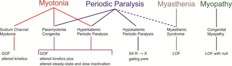
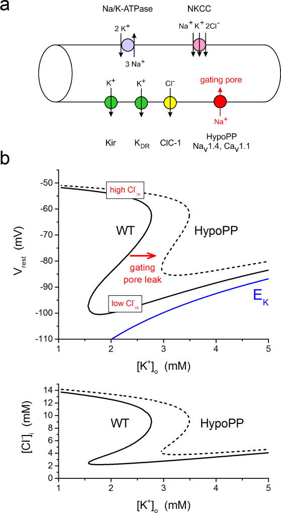
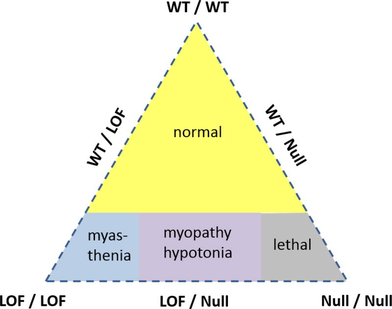
The NaV1.4 sodium channel is highly expressed in skeletal muscle, where it carries almost all of the inward Na+ current that generates the action potential, but is not present at significant levels in other tissues. Consequently, mutations of SCN4A encoding NaV1.4 produce pure skeletal muscle phenotypes that now include six allelic disorders: sodium channel myotonia, paramyotonia congenita, hyperkalemic periodic paralysis, hypokalemic periodic paralysis, congenital myasthenia, and congenital myopathy with hypotonia. Mutation-specific alternations of NaV1.4 function explain the mechanistic basis for the diverse phenotypes and identify opportunities for strategic intervention to modify the burden of disease.

Keywords: Channelopathy; Gating pore; Myotonia; NaV1.4; Periodic paralysis; Sodium channel.


Figures

Figure XX.1
Figure XX.1
Spectrum of clinical phenotypes, muscle diseases, and functional deficits for NaV1.4 channelopathies. Paramyotonia congenita and hyperkalemic periodic paralysis have considerable overlap in clinical features and NaV1.4 deficits. Sustained fluctuations in muscle strength may occur with myasthenia (dashed line), but the relation to periodic paralysis is uncertain. Abbreviations: GOF gain-of-function, LOF loss-of-function.
Figure XX.2
Figure XX.2
Impairment of fast inactivation for NaV1.4 mutations found in myotonia and HyperPP. (a) Cell-attached patch recordings from HEK cells show channel re-openings and prolonged open events for the two most commonly occurring HyperPP mutations (T704M and M1592V). Ensemble average (bottom) shows a small non-inactivating component (adapted from (Cannon and Strittmatter, 1993)) (b) Amplitude normalized whole-cell current shows a slower rate of inactivation for the SCM mutation F1705I (adapted from (Wu et al., 2005)). (c) Accelerated rate of recovery from fast inactivation for the PMC mutation T1313M (adapted from (Hayward et al., 1996)).
Figure XX.3
Figure XX.3
Model simulation of myotonia and periodic paralysis resulting from gain-of-function defects in NaV1.4. Top row shows simulated NaV1.4 mutant currents (blue lines) typical for SCM (middle) and HyperPP (right). A two-compartment model for skeletal muscle (Cannon et al., 1993a), to simulate the sarcolemma and the t-tubule including K+ accumulation, was used to simulate the response to simulated current injection. The simulated muscle normally fires a single action potential and then accommodates (left, note the difference in time scale compared to the top row). A reduced rate for onset of fast inactivation as in SCM (middle) gives rise to a sustained burst of myotonic discharges that persists after termination of the stimulus. A small persistent Na+ current to simulate HyperPP (right) also results in an initial myotonic burst, but then the membrane potential settles on an anomalously depolarized value, which inactivates the majority of NaV1.4 channels and renders the fiber refractory from a second stimulus pulse.
Figure XX.4
Figure XX.4
Gating pore currents in NaV1.4 HypoPP mutant channels. (a) Schematic representation of the NaV1.4 α subunit showing the location of missense mutations associated with muscle syndromes that include periodic paralysis (PMC, HyperPP, HypoPP). The HypoPP mutations are all at arginine residences in S4 segments and none are in domain IV. (b) Currents recorded from oocytes expressing wild type (WT, black) or R669H HypoPP mutant channels (red) in the presence of 1 µM TTX to block the NaV1.4 pore. The steady-state I–V relation shows inward rectification for R669H but not WT channels. (C) Inward rectification is consistent with a gating pore current resulting from a voltage-dependent anomalous conduction pathway that allows cation permeation only when the mutant residue of S4 is within the gating charge transfer center.
Figure XX.5
Figure XX.5
Simulated paradoxical depolarization in low K+ for a model HypoPP fiber. (a) Diagram of the pumps and transporters (top side) and ion channels (bottom side) to simulate the dependence of Vrest on extracellular K+. Arrows indicate the direction of net transport for specific ions when the fiber is at the normal Vrest. The gating pore is present in HypoPP fibers only, has a reversal potential near 0 mV, and in this simulation is carried primarily Na+ influx. (b) Phase plot of Vrest (top) and intracellular Cl (bottom) as the extracellular K+ was varied from 1 to 5 mM. At each set value of K+, the simulation searched for values of the membrane potential and internal Cl that simultaneously satisfied two constraints: that the sum of all ionic currents was zero (equilibrium point for Vm) and that the net Cl flux was also zero (mass balance for NKCC influx and ClC-1 efflux). For comparison, the Nernst potential for K+ is shown in blue. For a simulated WT fiber (solid line), Vrest hyperpolarizes as K+ is lowered from 5 mM until about 1.5 mM, at which point the fiber paradoxically depolarizes to −52 mV. This transition occurs because in very low K+ the Kir conductance can no longer balance the inward Cl current. The fiber depolarizes, which must be accompanied with an increase of intracellular Cl (bottom) because of the high resting Cl conductance. Addition of the gating pore current in a simulated HypoPP fiber (dashed line), shifts the relation to the right such that the paradoxical depolarization now occurs at 3 mM which is in the low physiologic range. The system has the possibility of multiple equilibrium potentials at a single value of external K+. For example, in the WT fiber this region of bistability for Vrest was with K+ between 1.5 and 2.75 mM. The internal [Cl] determines which value of Vrest will the fiber will settle upon (hyperpolarized for low Cl and depolarized for high Cl).
Figure XX.6
Figure XX.6
Diagram illustrating the relationship between wild-type (WT), loss-of-function (loss), and null alleles for NaV1.4 in determining the clinical phenotype. The loss-of-function refers to mutant NaV1.4 channels that express and conduct Na+ current, albeit with reduced amplitude or duration, as distinct from the complete absence of function with a null allele. (Reproduced from (Cannon, 2016)).

Similar articles

Cited by

References

    1. Aickin CC, Betz WJ, Harris GL. Intracellular chloride and the mechanism for its accumulation in rat lumbrical muscle. J Physiol. 1989;411:437–455. - PMC - PubMed
    1. Arnold WD, Feldman DH, Ramirez S, He L, Kassar D, Quick A, Klassen TL, Lara M, Nguyen J, Kissel JT, Lossin C, Maselli RA. Defective fast inactivation recovery of Nav 1.4 in congenital myasthenic syndrome. Ann Neurol. 2015;77:840–850. - PMC - PubMed
    1. Brancati F, Valente EM, Davies NP, Sarkozy A, Sweeney MG, LoMonaco M, Pizzuti A, Hanna MG, Dallapiccola B. Severe infantile hyperkalaemic periodic paralysis and paramyotonia congenita: broadening the clinical spectrum associated with the T704M mutation in SCN4A. J Neurol Neurosurg Psychiatry. 2003;74:1339–1341. - PMC - PubMed
    1. Brown GL, Harvey AM. Congenital myotonia in the goat. Brain. 1939;62:341–363.
    1. Caldwell JH, Campbell DT, Beam KG. Na channel distribution in vertebrate skeletal muscle. J Gen Physiol. 1986;87:907–932. - PMC - PubMed

Publication types

Substances

LinkOut - more resources