

Authentication of M14 melanoma cell line proves misidentification of MDA-MB-435 breast cancer cell line
- PMID: 28940260
- PMCID: PMC5762610
- DOI: 10.1002/ijc.31067
Authentication of M14 melanoma cell line proves misidentification of MDA-MB-435 breast cancer cell line
Abstract
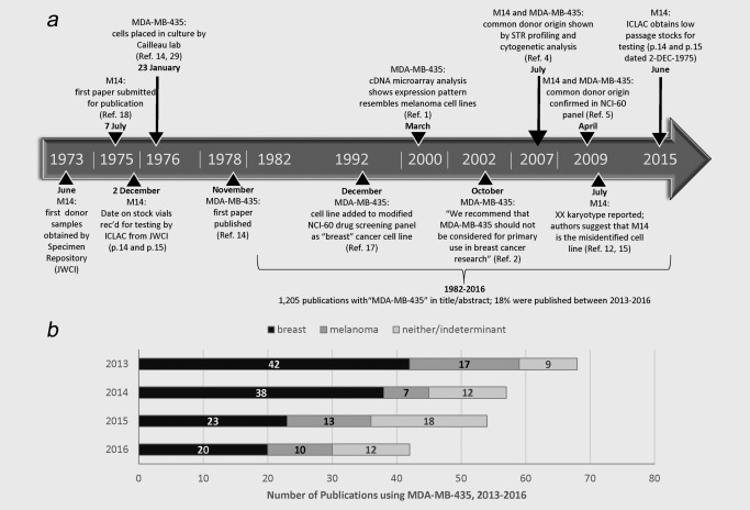
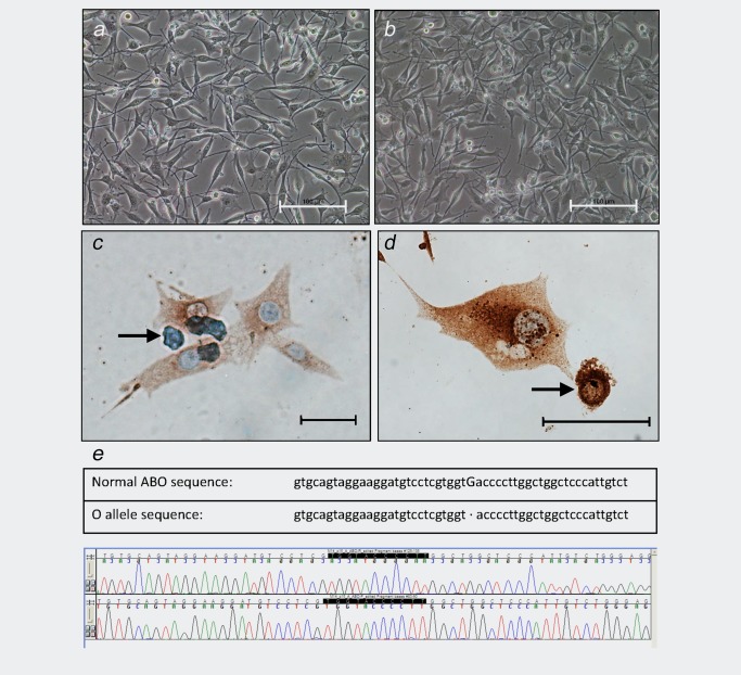
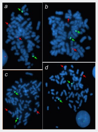
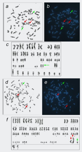
A variety of analytical approaches have indicated that melanoma cell line UCLA-SO-M14 (M14) and breast carcinoma cell line MDA-MB-435 originate from a common donor. This indicates that at some point in the past, one of these cell lines became misidentified, meaning that it ceased to correspond to the reported donor and instead became falsely identified (through cross-contamination or other means) as a cell line from a different donor. Initial studies concluded that MDA-MB-435 was the misidentified cell line and M14 was the authentic cell line, although contradictory evidence has been published, resulting in further confusion. To address this question, we obtained early samples of the melanoma cell line (M14), a lymphoblastoid cell line from the same donor (ML14), and donor serum preserved at the originator's institution. M14 samples were cryopreserved in December 1975, before MDA-MB-435 cells were established in culture. Through a series of molecular characterizations, including short tandem repeat (STR) profiling and cytogenetic analysis, we demonstrated that later samples of M14 and MDA-MB-435 correspond to samples of M14 frozen in 1975, to the lymphoblastoid cell line ML14, and to the melanoma donor's STR profile, sex and blood type. This work demonstrates conclusively that M14 is the authentic cell line and MDA-MB-435 is misidentified. With clear provenance information and authentication testing of early samples, it is possible to resolve debates regarding the origins of problematic cell lines that are widely used in cancer research.
Keywords: STR profiling; authentication; cross-contamination; human cell lines; misidentification.
© 2017 The Authors International Journal of Cancer published by John Wiley & Sons Ltd on behalf of UICC.
Figures
References
-
- Ross DT, Scherf U, Eisen MB, et al. Systematic variation in gene expression patterns in human cancer cell lines. Nat Genet 2000;24:227–35. - PubMed
-
- Rae JM, Ramus SJ, Waltham M, et al. Common origins of MDA‐MB‐435 cells from various sources with those shown to have melanoma properties. Clin Exp Metastasis 2004;21:543–52. - PubMed
-
- Rae JM, Creighton CJ, Meck JM, et al. MDA‐MB‐435 cells are derived from M14 melanoma cells–a loss for breast cancer, but a boon for melanoma research. Breast Cancer Res Treat 2007;104:13–9. - PubMed
Publication types
MeSH terms
Substances
Grants and funding
LinkOut - more resources
Full Text Sources
Other Literature Sources
Medical
Research Materials
Miscellaneous
