. 2017 Dec;26(12):2367-2380.
doi: 10.1002/pro.3304. Epub 2017 Oct 25.

The catalytic mechanism of cyclic GMP-AMP synthase (cGAS) and implications for innate immunity and inhibition

Affiliations

The catalytic mechanism of cyclic GMP-AMP synthase (cGAS) and implications for innate immunity and inhibition

Justin Hall et al. Protein Sci. 2017 Dec.

Abstract

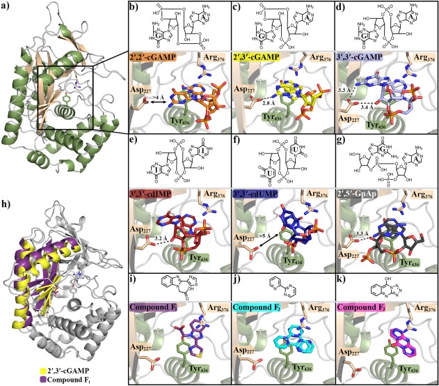
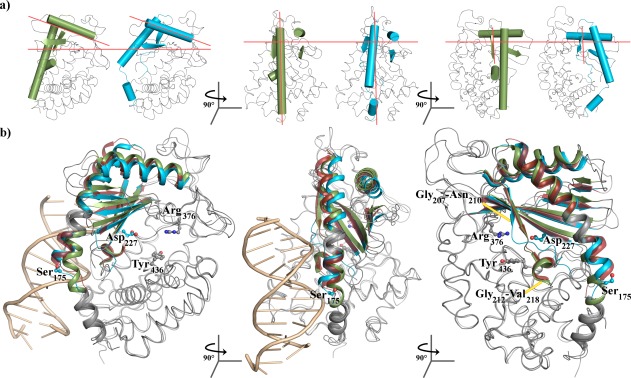
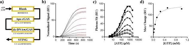
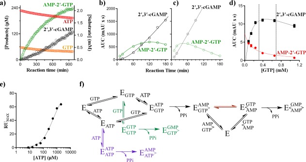
Cyclic GMP-AMP synthase (cGAS) is activated by ds-DNA binding to produce the secondary messenger 2',3'-cGAMP. cGAS is an important control point in the innate immune response; dysregulation of the cGAS pathway is linked to autoimmune diseases while targeted stimulation may be of benefit in immunoncology. We report here the structure of cGAS with dinucleotides and small molecule inhibitors, and kinetic studies of the cGAS mechanism. Our structural work supports the understanding of how ds-DNA activates cGAS, suggesting a site for small molecule binders that may cause cGAS activation at physiological ATP concentrations, and an apparent hotspot for inhibitor binding. Mechanistic studies of cGAS provide the first kinetic constants for 2',3'-cGAMP formation, and interestingly, describe a catalytic mechanism where 2',3'-cGAMP may be a minor product of cGAS compared with linear nucleotides.

Keywords: 2′,3′-cGAMP; OAS1; STING; cGAMP; cGAS; innate immunity.


Figures

Figure 1
Figure 1
Tyr436 and Arg376 form a binding site for aromatic rings. (a) cGAS161 bound to (b) 2′,2′‐cGAMP, (c) 2′,3′‐cGAMP, (d) 3′,3′‐cGAMP, (e) 3′,3′‐cdIMP, (f) 3′,3′‐cdUMP, and (g) 2′,5′‐GpAp. h) Changes between the inactive β‐sheet pose (compound F1 bound) and β‐pseudo active conformations (2′,3′‐cGAMP bound). cGAS161 bound to compound (i) F1, (j) F2, and (k) F3. Compounds structures are shown above protein structures; F2 and F3 are modeled with more than a single pose due to uncertainty in their electron density. Figure generated using Pymol with pdb structures 5VDO (cGAS161•2′,2′‐cGAMP), 5VDP (cGAS161•2′,3′‐cGAMP), 5VDT (cGAS161•3′,3′‐cGAMP), 5VDR (cGAS161•3′,3′‐cdIMP), 5VDS (cGAS161•3′,3′‐cdUMP), 5VDQ (cGAS161•2′,5′‐GpAp), 5VDW (cGAS161•F1), 5VDU (cGAS161•F2), and 5VDV (cGAS161•F3)28, 33, 34
Figure 2
Figure 2
cGAS is able to assume pseudo‐active states without binding to ds‐DNA. (a) cGAS in the α‐pseudo‐active (green) or β‐pseudo‐active conformation (blue). Orange lines are a guide for the eye corresponding to the approximate orientation of these same secondary structures in the ds‐DNA bound cGAS structure. (b) Overlay of cGAS bound to F1 (green) showing the α‐pseudo‐active conformation, 2′,3′‐cGAMP showing the β‐pseudo‐active conformation (blue), or ds‐DNA (red) showing the fully active conformation. In the third panel the ds‐DNA is forefront and has been removed for clarity. Images were generated using Pymol with pdb structures 5VDP (2′,3′‐cGAMP‐bound cGAS161), 5VDW (F1‐bound cGAS161), and 4KB6 (ds‐DNA‐, ATP‐, and GTP‐bound S. scrofa cGAS)
Figure 3
Figure 3
An SPR‐based enzymatic assay. (a) Schematic for SPR sensor layout. Individual channels are indicated by yellow boxes and the flow‐path is indicated by the black arrow. b) Representative data for normalized STING155–341 response over 900 s injections; data are for a fixed concentration of GTP (0.5 mM), variable ATP (3.9 μM to 2 mM over 10 points). The red lines show the fit of the data to a single exponential association from 600 to 900 s to determine the RU maxima. (c) Extrapolated RU maxima for seven fixed GTP concentration (16 μM to 1 mM) over variable ATP concentrations (3.9 μM to 2 mM). Solid lines show the fit of the data to equation (2) using a GTP‐concentration independent K M.ATP value with substrate inhibition observed at high nucleotide concentrations. (d) Calculated maximum response values from panel c were plotted as a function of the GTP concentration. The black line shows the fit of the data using the Michaelis‐Menten equation. Error bars of the fits for are within the size of the data markers in panels (c) and (d)
Figure 4
Figure 4
Linear homo‐ and hetero‐dinucleotides are the major products of cGAS. (a) NMR‐based full length cGAS reaction progress monitoring the loss of substrates (ATP and GTP, red and orange) and accumulation of AMP‐2′‐GTP (green) and 2′,3′‐cGAMP (black). HPLC‐based monitoring of AMP‐2′‐GTP (green) and 2′,3′‐cGAMP (black) from either b) cGAS161 or (c) full length cGAS. (d) Steady‐state rates for full length cGAS formation of 2′,3′‐cGAMP (black) or AMP‐3′‐ATP (red) at 1.1 mM ATP as a function of GTP; marked region represents approximate cellular GTP concentrations. (e) Maximum RU signal for STING binding to 2′,3′‐cGAMP at increasing concentrations of a fixed ratio (2:1) of ATP and GTP. The concentration of GTP is omitted on the x‐axis for simplicity. (f) Mechanism for the formation of homo‐ and hetero‐dinucleotides by cGAS (E); cGAS is shown after ds‐DNA activation; AMP‐2′‐GTP release or reorganization on‐enzyme (red path) are illustrated; schematic based on mechanism proposed by Gao et al19

Similar articles

Cited by

References

    1. Wang Q, Liu X, Zhou Q, Wang C (2015) Cytosolic sensing of aberrant DNA: arming STING on the endoplasmic reticulum. Expert Opin Ther Targets 19:1397–1409. - PubMed
    1. Schlee M, Hartmann G (2016) Discriminating self from non‐self in nucleic acid sensing. Nat Rev Immunol 16:566–580. - PMC - PubMed
    1. Junt T, Barchet W (2015) Translating nucleic acid‐sensing pathways into therapies. Nat Rev Immunol 15:529–544. - PubMed
    1. Jeremiah N, Neven B, Gentili M, Callebaut I, Maschalidi S, Stolzenberg MC, Goudin N, Fremond ML, Nitschke P, Molina TJ, Blanche S, Picard C, Rice GI, Crow YJ, Manel N, Fischer A, Bader‐Meunier B, Rieux‐Laucat F (2014) Inherited STING‐activating mutation underlies a familial inflammatory syndrome with lupus‐like manifestations. J Clin Invest 124:5516–5520. - PMC - PubMed
    1. Crow YJ (2011) Type I interferonopathies: a novel set of inborn errors of immunity. Ann NY Acad Sci 1238:91–98. - PubMed

LinkOut - more resources