. 2018 Jan;27(1):233-244.
doi: 10.1002/pro.3307. Epub 2017 Oct 10.

The human protein atlas: A spatial map of the human proteome

Affiliations

The human protein atlas: A spatial map of the human proteome

Peter J Thul et al. Protein Sci. 2018 Jan.

Abstract

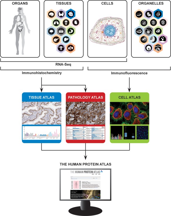
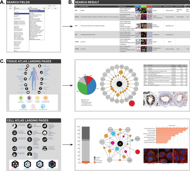
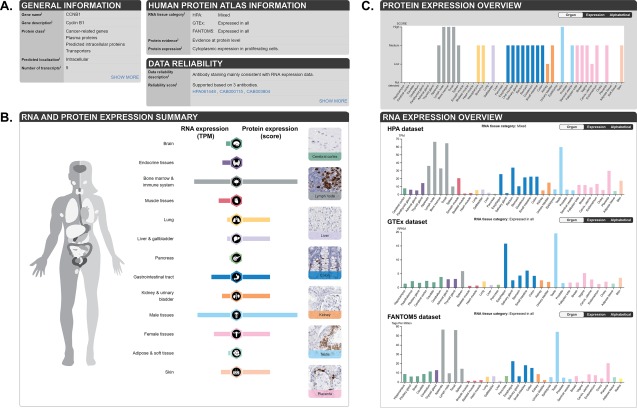
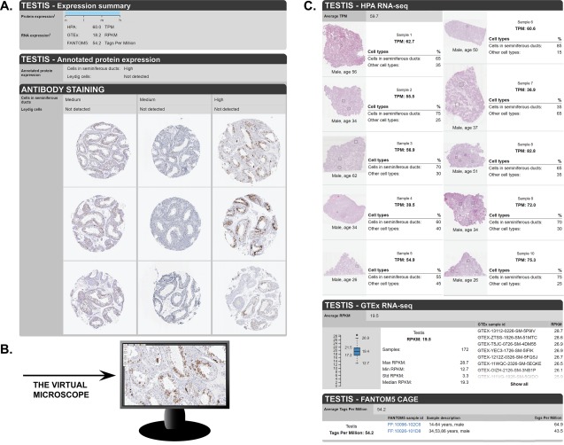
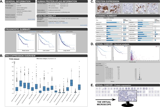
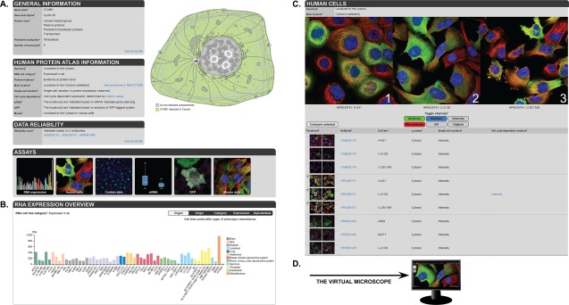
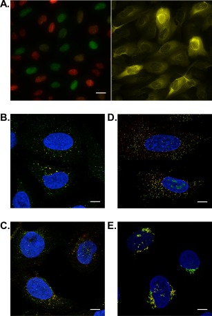
The correct spatial distribution of proteins is vital for their function and often mis-localization or ectopic expression leads to diseases. For more than a decade, the Human Protein Atlas (HPA) has constituted a valuable tool for researchers studying protein localization and expression in human tissues and cells. The centerpiece of the HPA is its unique antibody collection for mapping the entire human proteome by immunohistochemistry and immunocytochemistry. By these approaches, more than 10 million images showing protein expression patterns at a single-cell level were generated and are publicly available at www.proteinatlas.org. The antibody-based approach is combined with transcriptomics data for an overview of global expression profiles. The present article comprehensively describes the HPA database functions and how users can utilize it for their own research as well as discusses the future path of spatial proteomics.

Keywords: antibodies; immunofluorescence; immunohistochemistry; protein expression; single-cell; spatial proteomics; transcriptomics.


Figures

Figure 1
Figure 1
Schematic overview of the HPA. The HPA analyzes the human genome on different levels: in organs, tissues, cells, and organelles. Organs and tissues are stained using IHC, providing the basis for the Tissue Atlas and Pathology Atlas, while cells and organelles are analyzed with IF in the Cell Atlas. The proteomic analysis is combined with RNA‐Seq on the organ, tissue, and cellular level, and all data is freely accessible on the HPA web portal, www.proteinatlas.org.
Figure 2
Figure 2
Overview of different search strategies in the HPA. (A) Using the search fields allows for combining different criteria to a refined search for a particular group of proteins. (B) A search results in a list of genes meeting the criteria, with links to primary data in the different sub‐atlases. (C) Landing pages in the Tissue Atlas (top) and the Cell Atlas (bottom) contain numerous clickable charts, figures, images, and tables that allow for further investigation of certain proteins of interest.
Figure 3
Figure 3
Overview of a gene page in the Tissue Atlas. (A) On top of the page, general gene/protein information as well as the HPA data are summarized. (B) Summary of RNA and protein expression in 13 different groups of tissues, including examples of IHC stained tissues. (C) The plots depict protein expression levels in 44 non‐diseased tissues, as well as RNA expression levels from three different sources: the HPA (37 tissues), the GTEx consortium (31 tissues), and FANTOM5 (35 tissues).
Figure 4
Figure 4
Examples of a detailed tissue page in the Tissue Atlas. (A) On top of the page, a summary of protein and RNA expression levels in a particular tissue (here: testis) is shown. Below are annotated protein expression levels in the different cell types, as well as the primary antibody staining data for different antibodies, including three TMA cores per tissue and antibody. (B) Clicking on an image opens up an enlarged view that can be used like virtual microscope. (C) The samples used for analysis of RNA expression from three different datasets (HPA, GTEx, and FANTOM5) are shown, as well as individual values for each sample in the particular tissue.
Figure 5
Figure 5
Overview of a gene page in the Pathology Atlas. (A) On top of the page, general gene/protein information as well as the HPA data are summarized. Below, Kaplan‐Meier plots are shown for cancer types showing a significant prognostic association (here: renal, liver and lung cancer). (B) Box plots summarize the RNA expression levels across 17 different cancer types. (C) The protein expression section displays examples of IHC stained cancer tissues and the blue bars summarize cancer tissue staining pattern as well as protein expression of the corresponding non‐diseased tissue. (D) The detailed page shows survival analysis in a particular cancer type (here: renal cancer), as well as individual RNA expression values for each sample. (E) Primary antibody staining data for different antibodies are displayed. Clicking on an image opens up an enlarged view that can be used like virtual microscope.
Figure 6
Figure 6
Overview of a gene page in the Cell Atlas. (A) On top of the page, general gene/protein information as well as the HPA data are summarized. A schematic cell on the right highlights the sub‐cellular localization in green (here: cytosol). Miniature images show the different assays that were performed for this particular protein. (B) The plot depicts RNA expression levels in 56 analyzed cell lines. (C) Three high‐resolution IF images using different antibodies and cell lines can be selected and compared by clicking on the checkbox or by drag and drop. Single‐cell variations in gene expression are indicated and specified for variation in expression level (intensity) or spatial changes. (D) Clicking on an image opens up an enlarged view that can be used like a virtual microscope.
Figure 7
Figure 7
Examples of custom data in the Cell Atlas. (A) IF staining in the U‐2 OS FUCCI cell lines expressing Cdt1 (red) in G1 phase and Geminin (green) in S and G2 phases (left panel) reveals that CCNB1 expression peaks during S/G2 phase (right panel). Co‐localization studies using IF help to distinguish organelles comprised by the vesicle annotation or provide information about intraorganellar distribution. A yellow color indicates co‐localization revealing a localization of (B) ANKFY1 to endosomes (organelle marker EEA1), (C) TMEM192 to lysosomes (marker LAMP1), and (D) PEX14 to peroxisomes (marker ABCD3). (E) The Golgi apparatus is divided into several compartments. The Golgi‐associated protein ZFPL1 is only found in the cis‐Golgi compartment, as it co‐localizes with the marker cis‐Golgi protein GOLGA2. Scale bar 20 µm in (A) and 10 µm in (B–E).

Similar articles

Cited by

References

    1. Kim MS, Pinto SM, Getnet D, Nirujogi RS, Manda SS, Chaerkady R, Madugundu AK, Kelkar DS, Isserlin R, Jain S, Thomas JK, Muthusamy B, Leal‐Rojas P, Kumar P, Sahasrabuddhe NA, Balakrishnan L, Advani J, George B, Renuse S, Selvan LD, Patil AH, Nanjappa V, Radhakrishnan A, Prasad S, Subbannayya T, Raju R, Kumar M, Sreenivasamurthy SK, Marimuthu A, Sathe GJ, Chavan S, Datta KK, Subbannayya Y, Sahu A, Yelamanchi SD, Jayaram S, Rajagopalan P, Sharma J, Murthy KR, Syed N, Goel R, Khan AA, Ahmad S, Dey G, Mudgal K, Chatterjee A, Huang TC, Zhong J, Wu X, Shaw PG, Freed D, Zahari MS, Mukherjee KK, Shankar S, Mahadevan A, Lam H, Mitchell CJ, Shankar SK, Satishchandra P, Schroeder JT, Sirdeshmukh R, Maitra A, Leach SD, Drake CG, Halushka MK, Prasad TS, Hruban RH, Kerr CL, Bader GD, Iacobuzio‐Donahue CA, Gowda H, Pandey A (2014) A draft map of the human proteome. Nature 509:575–581. - PMC - PubMed
    1. Wilhelm M, Schlegl J, Hahne H, Gholami AM, Lieberenz M, Savitski MM, Ziegler E, Butzmann L, Gessulat S, Marx H, Mathieson T, Lemeer S, Schnatbaum K, Reimer U, Wenschuh H, Mollenhauer M, Slotta‐Huspenina J, Boese JH, Bantscheff M, Gerstmair A, Faerber F, Kuster B (2014) Mass‐spectrometry‐based draft of the human proteome. Nature 509:582. - PubMed
    1. Omenn GS, Lane L, Lundberg EK, Beavis RC, Overall CM, Deutsch EW (2016) Metrics for the Human Proteome Project 2016: progress on identifying and characterizing the human proteome, including post‐translational modifications. J Proteome Res 15:3951–3960. - PMC - PubMed
    1. Uhlén M, Fagerberg L, Hallström BM, Lindskog C, Oksvold P, Mardinoglu A, Sivertsson Å, Kampf C, Sjöstedt E, Asplund A, Olsson I, Edlund K, Lundberg E, Navani S, Szigyarto CA, Odeberg J, Djureinovic D, Takanen JO, Hober S, Alm T, Edqvist PH, Berling H, Tegel H, Mulder J, Rockberg J, Nilsson P, Schwenk JM, Hamsten M, von Feilitzen K, Forsberg M, Persson L, Johansson F, Zwahlen M, von Heijne G, Nielsen J, Pontén F (2015) Tissue‐based map of the human proteome. Science 347:6220. - PubMed
    1. Thul PJ, Åkesson L, Wiking M, Mahdessian D, Geladaki A, Ait Blal H, Alm T, Asplund A, Björk L, Breckels LM, Bäckström A, Danielsson F, Fagerberg L, Fall J, Gatto L, Gnann C, Hober S, Hjelmare M, Johansson F, Lee S, Lindskog C, Mulder J, Mulvey CM, Nilsson P, Oksvold P, Rockberg J, Schutten R, Schwenk JM, Sivertsson Å, Sjöstedt E, Skogs M, Stadler C, Sullivan DP, Tegel H, Winsnes C, Zhang C, Zwahlen M, Mardinoglu A, Pontén F, von Feilitzen K, Lilley KS, Uhlén M, Lundberg E (2017) A subcellular map of the human proteome. Science 356:eaal3321. - PubMed

Publication types