Review
. 2018 Oct 15;180(Pt B):515-525.
doi: 10.1016/j.neuroimage.2017.09.036. Epub 2017 Sep 21.

The behavioral and cognitive relevance of time-varying, dynamic changes in functional connectivity

Affiliations
Review

The behavioral and cognitive relevance of time-varying, dynamic changes in functional connectivity

Jessica R Cohen. Neuroimage. .

Abstract

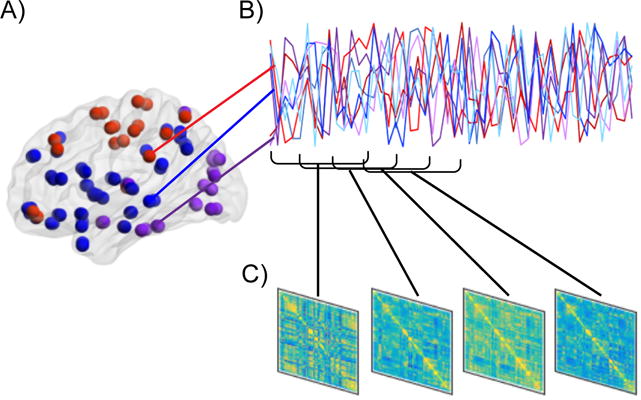
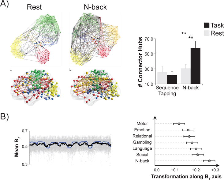
Recent advances in neuroimaging methods and analysis have led to an expanding body of research that investigates how large-scale brain network organization dynamically adapts to changes in one's environment, including both internal state changes and external stimulation. It is now possible to detect changes in functional connectivity that occur on the order of seconds, both during an unconstrained resting state and during the performance of constrained cognitive tasks. It is thought that these dynamic, time-varying changes in functional connectivity, often referred to as dynamic functional connectivity (dFC), include features that are relevant to behavior and cognition. This review summarizes four aspects of the nascent literature directly testing that assumption: 1) how changes in functional network organization on the order of task blocks relate to differences in task demands and to cognitive ability; 2) how differences in dFC variability between different contexts relate to cognitive demands and behavioral performance; 3) how ongoing fluctuations in dFC impact perception and attention; and 4) how different patterns of dFC correspond to individual differences in cognition. The review ends by discussing promising directions for future research in this field. First, it comments on how dFC analyses can help to elucidate the mechanisms of healthy cognition. Next, it describes how dFC processes may be disrupted in disease, and how probing such dysfunction can increase understanding of neural etiology, as well as behavioral and cognitive impairments, observed in psychiatric and neurologic populations. Last, it considers the potential for computational models to uncover neuronal mechanisms of dFC, and how both healthy cognition and disease emerge from network dynamics.

Keywords: Cognition; Dynamic functional connectivity; Individual differences; Network dynamics; Resting state; Time-varying.


Figures

Figure 1
Figure 1. Steps to conduct a dFC analysis
A) First, the brain is parcellated into nodes, which can consist of anatomical or functional regions of interest, or components derived from a data-driven method such as independent component analysis. B) Second, the time-series across all pairs of nodes are related to each other, often by computing correlations or coherence, but other methods such as co-activation patterns or temporal derivatives can be used as well. Commonly, this is repeated within pre-specified and overlapping “windows” of fixed length (as pictured), but novel methods that do not require the assumptions of sliding window approaches can also be utilized, such as dynamical conditional correlations (Lindquist et al., 2014), multiplication of temporal derivatives (Shine et al., 2015) or co-activation patterns (Liu and Duyn, 2013). C) Last, individual connectivity matrices are computed for each window. Once multiple FC matrices are computed for each time-series, dFC analyses quantifying how the matrices differ from each other can be conducted.
Figure 2
Figure 2. DFC analyses complement and extend static FC analyses
A) Using static FC methods, whole-brain functional network organization was found to reconfigure between rest and an n-back task that probed working memory (Cohen and D’Esposito, 2016). In the left panel each color represents a network, each colored line represents a within-network edge, and each black line represents a between-network edge. On the top, nodes are depicted based on connections; nodes with more shared connections are closer together. On the bottom, nodes are depicted in brain space; each circle corresponds to the coordinates of the center of each node. Note that there is greater integration across distinct networks during the n-back task as compared to rest. In the right panel, the number of connector hub nodes is compared across rest, n-back, and during a sequence tapping task that probed motor execution. Connector hubs are nodes with high inter-network connectivity. The number of connector hubs did not change during sequence tapping as compared to rest, but it increased during the n-back task as compared to rest. Figure adapted with permission from Cohen and D’Esposito (2016). **p < .01. B) Using dFC methods, participation coefficient (BT) was found to fluctuate as current task changed (Shine et al., 2016a). Participation coefficient measures how connected a node is across networks; connector nodes are defined as nodes with high participation coefficients. The left panel demonstrates that using dFC analyses, average participation coefficient (thick black line; individual participant data plotted in gray) varied along with task blocks (task regressors plotted in blue). The right panel demonstrates the extent to which whole-brain FC profiles shifted toward a more integrated brain state (high BT) during different tasks as compared to rest. Consistent with Cohen and D’Esposito (2016), during motor task performance the extent of integration was most similar that during rest, while during n-back performance the extent of integration was much stronger. Figure adapted with permission from Shine et al. (2016a).

References

    1. Bargmann CI, Marder E. From the connectome to brain function. Nature Methods. 2013;10:483–490. - PubMed
    1. Barttfeld P, Uhrig L, Sitt JD, Sigman M, Jarraya B, Dehaene S. Signature of consciousness in the dynamics of resting-state brain activity. Proceedings of the National Academy of Sciences of the United States of America. 2015;112:887–892. - PMC - PubMed
    1. Bassett DS, Yang M, Wymbs NF, Grafton ST. Learning-induced autonomy of sensorimotor systems. Nature Neuroscience. 2015;18:744–751. - PMC - PubMed
    1. Braun U, Schäfer A, Walter H, Erk S, Romanczuk-Seiferth N, Haddad L, Schweiger JI, Grimm O, Heinz A, Tost H, Meyer-Lindenberg A, Bassett DS. Dynamic reconfiguration of frontal brain networks during executive cognition in humans. Proceedings of the National Academy of Sciences of the United States of America. 2015;112:11678–11683. - PMC - PubMed
    1. Breakspear M. Dynamic models of large-scale brain activity. Nature Neuroscience. 2017;20:340–352. - PubMed

Publication types