. 2017 Nov;128(11):2165-2178.
doi: 10.1016/j.clinph.2017.08.001. Epub 2017 Sep 5.

Pallidal low β-low γ phase-amplitude coupling inversely correlates with Parkinson disease symptoms

Affiliations

Pallidal low β-low γ phase-amplitude coupling inversely correlates with Parkinson disease symptoms

Christos Tsiokos et al. Clin Neurophysiol. 2017 Nov.

Abstract

Objective: Recent discoveries suggest that it is most likely the coupling of β oscillations (13-30Hz) and not merely their power that relates to Parkinson disease (PD) pathophysiology.

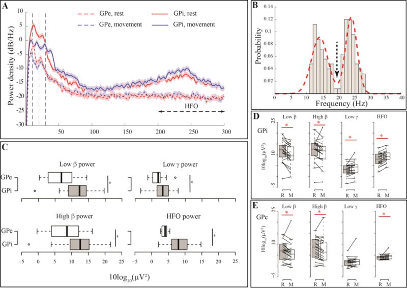
Methods: We analyzed power and phase amplitude coupling (PAC) in local field potentials (LFP) recorded from Pallidum after placement of deep brain stimulation (DBS) leads in nineteen PD patients and three patients with dystonia.

Results: Within GPi, we identified PAC between phase of β and amplitude of high frequency oscillations (200-300Hz) and distinct β-low γ (40-80Hz) PAC both modulated by contralateral movement. Resting β-low γ PAC, also present in dystonia patients, inversely correlated with severity of rigidity and bradykinesia (R=-0.44, P=0.028). These findings were specific to the low β band, suggesting a differential role for the two β sub-bands.

Conclusions: PAC is present across distinct frequency bands within the GPi. Given the presence of low β-low γ PAC in dystonia and the inverse correlation with symptom severity, we propose that this PAC may be a normal pallidal signal.

Significance: This study provides new evidence on the pathophysiological contribution of local pallidal coupling and suggests similar and distinct patterns of coupling within GPi and STN in PD.

Keywords: Basal ganglia; Deep brain stimulation; Parkinson disease; Phase-amplitude coupling; β oscillations.


Conflict of interest statement

Conflict of Interest

None of the authors have potential conflicts of interest to be disclosed.

Figures

Figure 1
Figure 1. DBS lead and bipolar contact pairs in the Globus pallidus. (A) Gross anatomy of globus pallidus and approximate location of DBS lead
The DBS lead was targeted to motor (ventral posterolateral) GPi using image-guided targeting, 2–4 mm anterior, 19–24 mm lateral and 4–6 mm inferior to the mid-commissural point and covers the spanning globus pallidus internus (GPi) and globus pallidus externus (GPe). (B–C) Co-registration of post-operative CT scan and Pre-operative T1-weighted structural MRI shows average location of bipolar pairs for the cohort in the standard space (MNI152) and confirms that for both hemispheres contact pair 0–1 (red) is located in the GPi and contact pair 2–3 is located in thhe GPe (blue).
Figure 2
Figure 2. Pallidal power, its spatial distribution and movement modulation
(A) Example PSD at bipolar channels (0–1 and 2–3) for one subject during rest and movemen, showing two prominent spectral peaks in low and high β frequency range. Movement causes β power suppression. (B) Histogram of spectral peaks in β range for all the PSDs investigated from channel pair (0–1) in all subjects. The arrow shows the local minima between two modes of the probability distribution which was selected as the boundary between low and high β sub-bands for further analyses. (C) Boxplots showing band power differences between GPi (0–1) and GPe (2–3) across the cohort, (*) indicates statistically significant difference between the pairs examined. (D–E) Effect of movement on different frequency bands for the 19 GPi (D) and GPe (E) nuclei contralateral to the movement body side.
Figure 3
Figure 3. Pallidal PAC, its spatial distribution and movement modulation
(A) Sample PAC comodulograms for one example subject at GPi (0–1) and GPe (2–3) during rest and movement. White circles show statistically significant regions after correction for multiple comparisons using FDR. (B) Kernel density estimates for distribution of (left) phase encoding and (right) amplitude frequencies for maximal coupling in the β-low γ and β-HFO PAC showing for majority of recordings, peak PAC was observed at low β phase further emphasizing the local role of pallidal low β. (C) boxplots showing β-low γ and β-HFO PAC are stronger at GPi (0–1) relative to the GPe (2–3) (D–E) Movement modulation of pallidal β-low γ and β-HFO PAC in GPi (D) and GPe (E) (N= 19, contralateral to the movement body side). (*) indicates statistically significant difference between the pairs examined.
Figure 4
Figure 4. Clinical correlates of Pallidal power and PAC during rest and their movement modulation
(A) Significant clinical correlation during rest between low β-low γ PAC (top row) and high β-low γ PAC (bottom row) and hemibody-UPDRS (left column) and sum of scores for rigidity and bradykinesia (right column) [N = 38]. (B) Significant clinical correlation with movement modulation [movement:rest] between low γ power ratio (top row) and low β-low γ PAC ratio (bottom row) and hemibody-UPDRS (left column) and sum of scores for rigidity and bradykinesia (right column) [N = 19].
Figure 5
Figure 5. Pallidal low β-low γ PAC during movement versus hemibody UPDRS
Subjects with different degrees of disease severity suppress their contralateral low β-low γ PAC during movement to similar values. (Black arrow indicates one outlier subject whose movement low β-low γ PAC was relatively higher than the rest of the cohort and was excluded from the clinical correlation analysis)

References

    1. Andersson JLR, Jenkinson M, Smith S. Non-linear registration aka Spatial normalisation. FMRIB Technial Report TR07JA2. 2007;22 In Pract.
    1. Androulidakis AG, Kuhn AA, Chen CC, Blomstedt P, Kempf F, Kupsch A, et al. Dopaminergic therapy promotes lateralized motor activity in the subthalamic area in Parkinson’s disease. Brain. 2007;130:457–68. doi: 10.1093/brain/awl358. - DOI - PubMed
    1. Aru JJ, Aru JJ, Priesemann V, Wibral M, Lana L, Pipa G, et al. Untangling cross-frequency coupling in neuroscience. Curr Opin Neurobiol. 2015;31:51–61. doi: 10.1016/j.conb.2014.08.002. - DOI - PubMed
    1. Benjamini Y, Drai D, Elmer G, Kafkafi N, Golani I. Controlling the false discovery rate in behavior genetics research. Behav Brain Res. 2001;125:279–84. - PubMed
    1. Brittain J-SS, Brown P. Oscillations and the basal ganglia: motor control and beyond. Neuroimage. 2014;85:637–47. doi: 10.1016/j.neuroimage.2013.05.084. - DOI - PMC - PubMed

Publication types