Review
. 2018 Jan:67:109-127.
doi: 10.1016/j.jbior.2017.09.002. Epub 2017 Sep 6.

Molecular basis of the human ribosomopathy Shwachman-Diamond syndrome

Affiliations
Review

Molecular basis of the human ribosomopathy Shwachman-Diamond syndrome

Alan J Warren. Adv Biol Regul. 2018 Jan.

Abstract

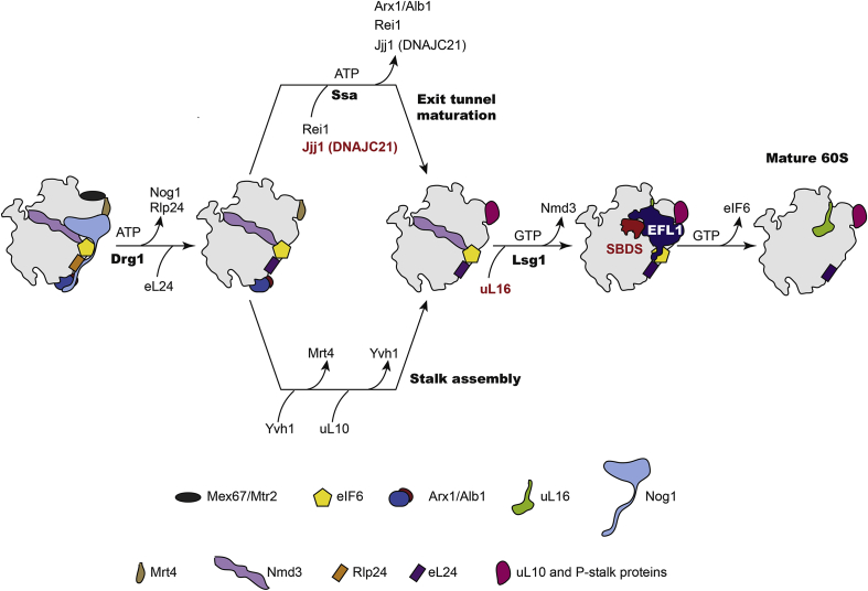
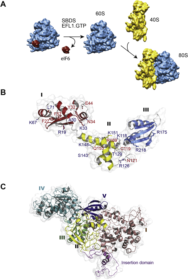
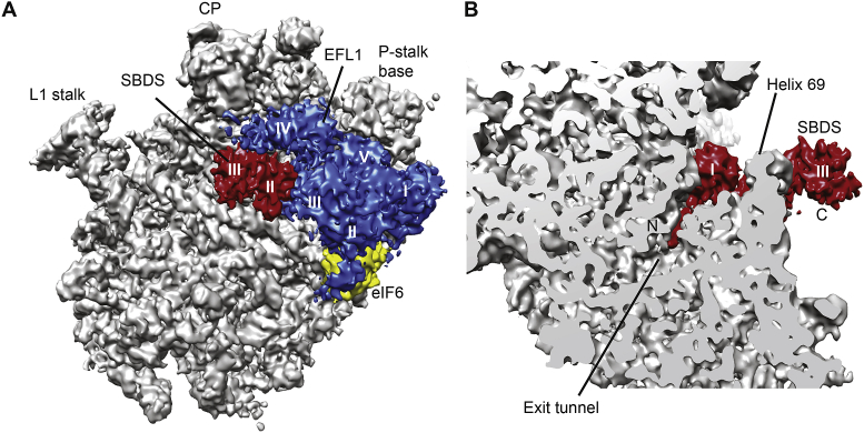
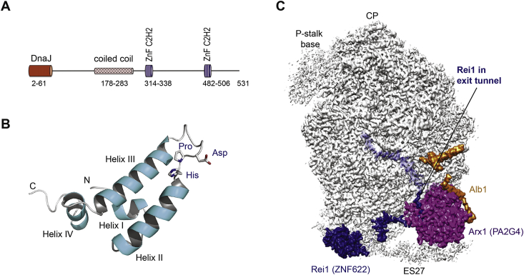
Mutations that target the ubiquitous process of ribosome assembly paradoxically cause diverse tissue-specific disorders (ribosomopathies) that are often associated with an increased risk of cancer. Ribosomes are the essential macromolecular machines that read the genetic code in all cells in all kingdoms of life. Following pre-assembly in the nucleus, precursors of the large 60S and small 40S ribosomal subunits are exported to the cytoplasm where the final steps in maturation are completed. Here, I review the recent insights into the conserved mechanisms of ribosome assembly that have come from functional characterisation of the genes mutated in human ribosomopathies. In particular, recent advances in cryo-electron microscopy, coupled with genetic, biochemical and prior structural data, have revealed that the SBDS protein that is deficient in the inherited leukaemia predisposition disorder Shwachman-Diamond syndrome couples the final step in cytoplasmic 60S ribosomal subunit maturation to a quality control assessment of the structural and functional integrity of the nascent particle. Thus, study of this fascinating disorder is providing remarkable insights into how the large ribosomal subunit is functionally activated in the cytoplasm to enter the actively translating pool of ribosomes.

Keywords: DNAJC21; Myelodysplastic syndromes; Ribosome; SBDS; Shwachman-Diamond syndrome; eIF6.


Figures

Fig. 1
Fig. 1
Schematic overview of cytoplasmic 60S subunit assembly. The AAA-ATPase Drg1 promotes release of the GTPase Nog1 and the exchange of Rlp24 for Rpl24 that in turn recruits Rei1. The J-protein Jjj1 (human DNAJC21) cooperates with the ATP-dependent Hsp70 chaperone SSa to release the export receptor Arx1 that lies close to the polypeptide exit tunnel. In a parallel pathway, Yvh1 facilitates the exchange of Mrt4 with P0 to assemble the ribosome stalk. Recruitment of uL16 in concert with the GTPase Lsg1 promotes release of the export adaptor Nmd3. Finally, release of eIF6 from the subunit joining face by SBDS and the EFL1 GTPase licences entry of the mature subunit into the pool of actively translating ribosomes. Proteins mutated in human disease are highlighted in red. DNAJC21 and SBDS are mutated in Shwachman-Diamond syndrome; uL16 is mutated in paediatric T-ALL.
Fig. 2
Fig. 2
SBDS and the EFL1 GTPase cooperate to catalyse eIF6 eviction. (A) Schematic showing that SBDS cooperates with the GTPase EFL1 in the cytoplasm to catalyse release of the anti-association factor eIF6 from the 60S subunit joining face to allow subunit joining and the formation of translation-competent 80S ribosomes. 40S subunit is coloured yellow, 60S subunit is cyan, eIF6 is red. (B) Ribbon representation of the human SBDS NMR structure. Domain I is coloured red, domain II is yellow, domain III is blue and loops are coloured grey. SDS-associated mutations modify surface epitopes (red text) or affect protein stability (blue text). (C) Ribbon representation of the atomic model of human EFL1 (Weis et al., 2015), with domain I coloured violet, domain II yellow, domain III green, domain IV cyan and domain V blue.
Fig. 3
Fig. 3
SBDS and EFL1 proofread the structural and functional integrity of the nascent 60S subunit. (A) Crown view and (B) transverse section of the cryo-EM maps of the 60S-eIF6-SBDS-EFL1 complex. The 60S subunit is coloured grey, SBDS is red, EFL1 is blue and eIF6 is yellow. SBDS domains II and III and EFL1 domains I-V are shown. CP, central protruberance.
Fig. 4
Fig. 4
The J-protein DNAJC21 facilitates release of the export receptor Arx1 (human PA2G4) from the pre-60S ribosomal subunit. (A) Schematic representation of the domain structure of human DNAJC21 (NP_001335349). ZnF, zinc finger. (B) Ribbon representation of the NMR structure of the J-domain (residues 2–108) from E. coli DnaJ protein (pdb 1xbl) (Pellecchia et al., 1996). J-domains comprise four α-helices. The invariant His, Pro, Asp (HPD) tripeptide in the loop between helices II and III is critical to stimulate the ATPase activity and in vivo function of its cognate cochaperone heat shock 70 kDa protein. (C) Visualising the 60S-bound assembly factors Arx1 (human PA2G4), Rei1 (human ZNF622) and Alb1 near the polypeptide exit tunnel (pdb 5APO) (Greber et al., 2012). The Arx1 protein shields the polypeptide exit tunnel, which is deeply probed by the C-terminus of the Rei1 protein. Alb1 likely modulates the affinity of Arx1 binding to the 60S subunit.

References

    1. Ajore R., Raiser D., McConkey M., Joud M., Boidol B., Mar B., Saksena G., Weinstock D.M., Armstrong S., Ellis S.R., Ebert B.L., Nilsson B. Deletion of ribosomal protein genes is a common vulnerability in human cancer, especially in concert with TP53 mutations. EMBO Mol. Med. 2017;9(4):498–507. - PMC - PubMed
    1. Armistead J., Khatkar S., Meyer B., Mark B.L., Patel N., Coghlan G., Lamont R.E., Liu S., Wiechert J., Cattini P.A., Koetter P., Wrogemann K., Greenberg C.R., Entian K.D., Zelinski T., Triggs-Raine B. Mutation of a gene essential for ribosome biogenesis, EMG1, causes Bowen-Conradi syndrome. Am. J. Hum. Genet. 2009;84(6):728–739. - PMC - PubMed
    1. Austin K.M., Gupta M.L., Coats S.A., Tulpule A., Mostoslavsky G., Balazs A.B., Mulligan R.C., Daley G., Pellman D., Shimamura A. Mitotic spindle destabilization and genomic instability in Shwachman-Diamond syndrome. J. Clin. Invest. 2008;118(4):1511–1518. - PMC - PubMed
    1. Austin K.M., Leary R.J., Shimamura A. The Shwachman-Diamond SBDS protein localizes to the nucleolus. Blood. 2005;106(4):1253–1258. - PMC - PubMed
    1. Ball H.L., Zhang B., Riches J.J., Gandhi R., Li J., Rommens J.M., Myers J.S. Shwachman-Bodian Diamond syndrome is a multi-functional protein implicated in cellular stress responses. Hum. Mol. Genet. 2009;18(19):3684–3695. - PMC - PubMed

Publication types

MeSH terms