. 2017 Oct 9;27(19):2984-2990.e3.
doi: 10.1016/j.cub.2017.08.014. Epub 2017 Sep 21.

The Jellyfish Cassiopea Exhibits a Sleep-like State

Affiliations

The Jellyfish Cassiopea Exhibits a Sleep-like State

Ravi D Nath et al. Curr Biol. .

Abstract

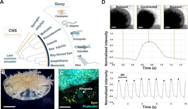
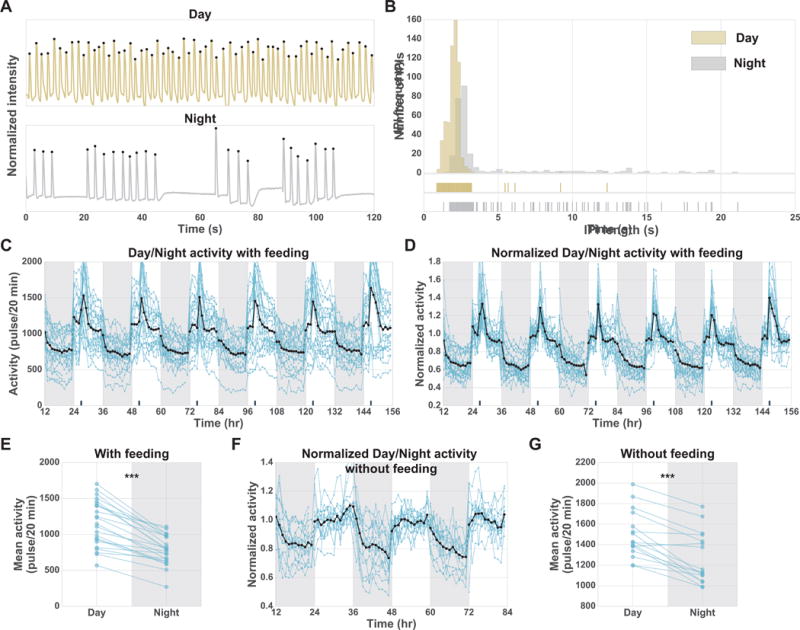
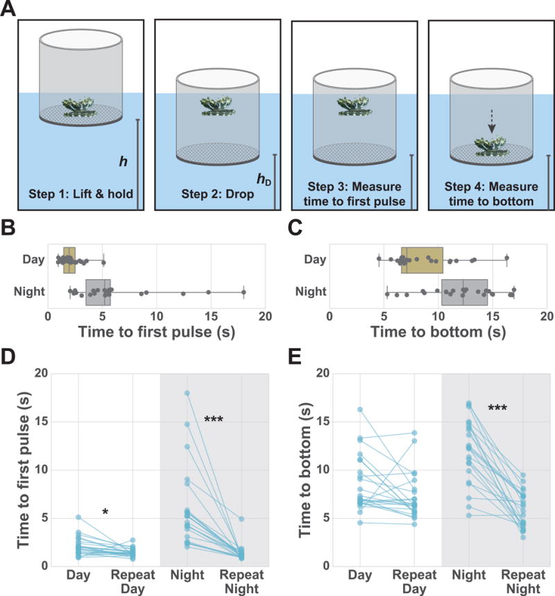
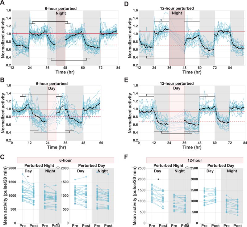
Do all animals sleep? Sleep has been observed in many vertebrates, and there is a growing body of evidence for sleep-like states in arthropods and nematodes [1-5]. Here we show that sleep is also present in Cnidaria [6-8], an earlier-branching metazoan lineage. Cnidaria and Ctenophora are the first metazoan phyla to evolve tissue-level organization and differentiated cell types, such as neurons and muscle [9-15]. In Cnidaria, neurons are organized into a non-centralized radially symmetric nerve net [11, 13, 15-17] that nevertheless shares fundamental properties with the vertebrate nervous system: action potentials, synaptic transmission, neuropeptides, and neurotransmitters [15-20]. It was reported that cnidarian soft corals [21] and box jellyfish [22, 23] exhibit periods of quiescence, a pre-requisite for sleep-like states, prompting us to ask whether sleep is present in Cnidaria. Within Cnidaria, the upside-down jellyfish Cassiopea spp. displays a quantifiable pulsing behavior, allowing us to perform long-term behavioral tracking. Monitoring of Cassiopea pulsing activity for consecutive days and nights revealed behavioral quiescence at night that is rapidly reversible, as well as a delayed response to stimulation in the quiescent state. When deprived of nighttime quiescence, Cassiopea exhibited decreased activity and reduced responsiveness to a sensory stimulus during the subsequent day, consistent with homeostatic regulation of the quiescent state. Together, these results indicate that Cassiopea has a sleep-like state, supporting the hypothesis that sleep arose early in the metazoan lineage, prior to the emergence of a centralized nervous system.

Keywords: Cassiopea; Cnidaria; evolution of sleep; jellyfish; sleep.


Figures

Figure 1
Figure 1. The pulsing behavior of the upside-down jellyfish, Cassiopea spp., is trackable
(A) Phylogenetic tree schematic highlighting animals in which sleep behavior has been described, the presence of neurons (tan), and the emergence of a centralized nervous system (dark blue). See boxed key. (B) An image of Cassiopea. (C) Higher magnification view of Cassiopea with labeled actin-rich muscle (phalloidin stain; cyan), autofluorescent Symbiodinium (yellow), and a rhopalia, the sensory organ that controls pulsing, which is free of Symbiodinium. (D) As Cassiopea pulse the relaxation and contraction of the bell causes a corresponding change in average pixel intensity. Pulsing behavior was tracked by measuring this change in pixel intensity within the region of interest. (top) Representative frames and corresponding normalized pixel intensities for one pulse event. The local maxima in the pulse-trace was used to count pulse events. (bottom) A 10-second recording of one jellyfish shows multiple pulsing events. The inter-pulse interval (IPI) was calculated as the time between the maxima. See Figure S1, Figure S2, Movie S1.
Figure 2
Figure 2. Continuous tracking of Cassiopea reveals pulsing quiescence at night
(A) Pulsing-traces for individual jellyfish during day and night over 120 s. (B) The distribution of IPI length for a 12-hour day and a 12-hour night for the same jellyfish shown in A. Tick marks below the distribution show each IPI length during the day and night. This highlights the long-pause events, which are more common at night (Figure S3A; Data S1). (C-G) Each blue line corresponds to a single jellyfish. The black line indicates the mean activity of all jellyfish. Dark gray shading indicates night periods. Dark tick marks on the x-axis indicate time of feeding. (C) Baseline activity (pulses/20 min) of 23 jellyfish tracked for six days from four laboratory replicates. (D) Normalized baseline activity for jellyfish shown in C, where each jellyfish is normalized by their mean day activity. (E) Mean day activity versus mean night activity for each jellyfish over the six-day experiment shown in C. Two-sided paired t-test, day versus night, P = 6×10⁻⁹. (F) Normalized baseline activity without feeding of 16 jellyfish tracked over three days from two laboratory replicates, where each jellyfish is normalized by its mean day activity. (G) Mean day activity versus mean night activity for each jellyfish over the three-day experiment shown in F. Two-sided paired t-test, day versus night, P =10−5. ***P<10−3. See Figure S3.
Figure 3
Figure 3. Cassiopea show reduced responsiveness to a sensory stimulus at night
(A) Schematic of experiment to test sensory responsiveness. Jellyfish were lifted and held at a fixed height (hL) and then dropped to a fixed height (hD). hL and hD were kept constant throughout experiments. Boxplots of time to first pulse after drop (B) for 23 jellyfish and time to reach bottom after drop (C) for 23 jellyfish during the day and night. Dots represent individual jellyfish collected from two laboratory replicates. Two-sided unpaired t-test, day versus night, (B) P < 10−4 and (C) P = 5×10⁻⁴. (D) Time to first pulse after initial drop and after perturbation for both day and night for 23 jellyfish. (E) Time to reach bottom after initial drop and after perturbation for both day and night for 23 jellyfish. Two-way analysis of variance (ANOVA) for data shown in D and E, followed by post-hoc comparisons between experimental groups using Bonferroni posttest (*P<5×10⁻², ***P<10−3). For the time to first pulse, two-sided unpaired t-test (B) and two-way ANOVA (D) were performed after log-transformation (Star Methods).
Figure 4
Figure 4. Homeostatic rebound in Cassiopea
Each blue line corresponds to a single jellyfish. The black line indicates the mean activity of all jellyfish. Dark gray shading indicates night periods. Maroon shading indicates perturbation periods with 10 s water pulses every 20 min. Jellyfish were exposed to different perturbation lengths (6 or 12 hours) at different times (day or night). The normalized activity of all jellyfish tracked over multiple days is plotted. Maroon horizontal lines show the mean activity of pre-perturbation day (solid) and pre-perturbation night (dashed). (A) Perturbation of 30 jellyfish for the last 6 hours of the night. (B) Perturbation of 26 jellyfish for the first 6 hours of the day. (C) Mean day and night activity pre- and post-perturbation for experiments shown in A and B. (D) Perturbation of 16 jellyfish for an entire 12-hour night. (E) Perturbation of 16 jellyfish for an entire 12-hour day. (F) Mean day and night activity pre- and post-perturbation for experiments shown in D and E. Black-horizontal lines in A, B, D, and E indicate the windows of time used for calculating pre- and post-perturbation means shown in C and F for both the night (bottom lines) and day (top lines). For the 6-hour experiments we compared the first 4 hours of the post-perturbation day to the equivalent time pre-perturbation, and also compared the first 6 hours of post-perturbation night to the equivalent time pre-perturbation. For the 12-hour experiments we compared the full 12-hour days and nights pre- and post-perturbation. Two-way ANOVA followed by post-hoc comparisons between experimental groups using Bonferroni posttest (*P<5×10⁻²). Both day and night 6-hour perturbation experiments include data from four laboratory replicates. Both day and night 12-hour perturbation experiments include data from two laboratory replicates. See Figure S4, Movie S2.

Comment in

References

    1. Hendricks JC, Finn SM, Panckeri KA, Chavkin J, Williams JA, Sehgal A, Pack AI. Rest in Drosophila is a sleep-like state. Neuron. 2000;25:129–138. - PubMed
    1. Shaw PJ, Cirelli C, Greenspan RJ, Tononi G. Correlates of sleep and waking in Drosophila melanogaster. Science. 2000;287:1834–1837. - PubMed
    1. Raizen DM, Zimmerman JE, Maycock MH, Ta UD, You YJ, Sundaram MV, Pack AI. Lethargus is a Caenorhabditis elegans sleep-like state. Nature. 2008;451:569–572. - PubMed
    1. Hill AJ, Mansfield R, Lopez JMNG, Raizen DM, Van Buskirk C. Cellular stress induces a protective sleep-like state in C. elegans. Curr Biol. 2014;24:2399–2405. - PMC - PubMed
    1. Trojanowski NF, Raizen DM. Call it worm sleep. Trends in Neurosciences. 2016;39:54–62. - PMC - PubMed