. 2017 Oct 23;43(2):227-239.e5.
doi: 10.1016/j.devcel.2017.08.021. Epub 2017 Sep 21.

CSNK1a1 Regulates PRMT1 to Maintain the Progenitor State in Self-Renewing Somatic Tissue

Affiliations

CSNK1a1 Regulates PRMT1 to Maintain the Progenitor State in Self-Renewing Somatic Tissue

Xiaomin Bao et al. Dev Cell. .

Abstract

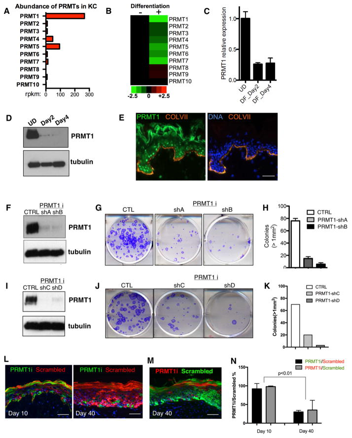
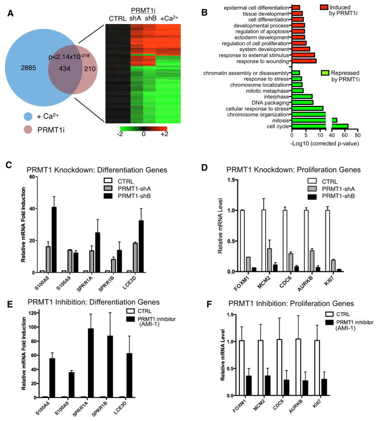
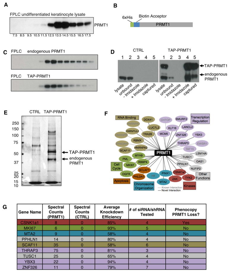
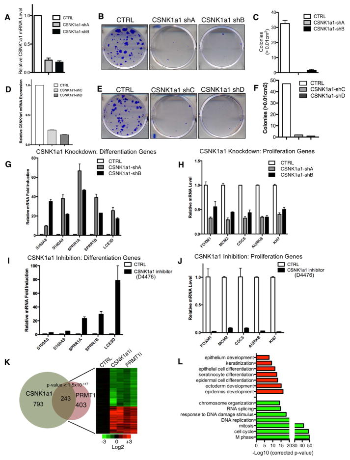
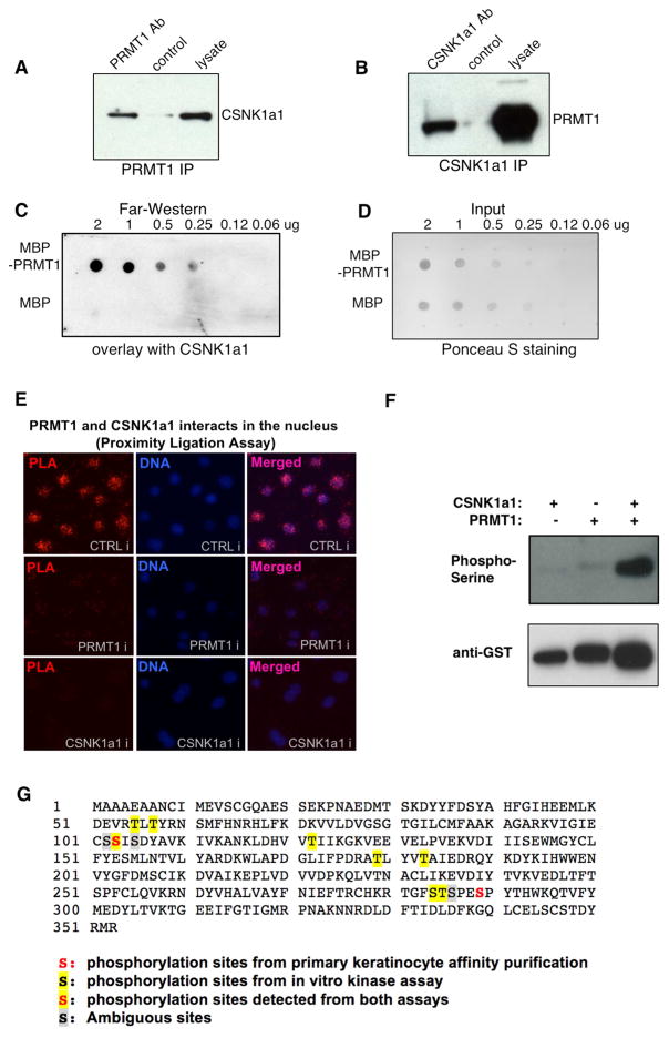
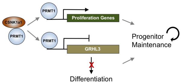
Somatic progenitors sustain tissue self-renewal while suppressing premature differentiation. Protein arginine methyltransferases (PRMTs) affect many processes; however, their role in progenitor function is incompletely understood. PRMT1 was found to be the most highly expressed PRMT in epidermal progenitors and the most downregulated PRMT during differentiation. In targeted mouse knockouts and in long-term regenerated human mosaic epidermis in vivo, epidermal PRMT1 loss abolished progenitor self-renewal and led to premature differentiation. Mass spectrometry of the PRMT1 protein interactome identified the CSNK1a1 kinase, which also proved essential for progenitor maintenance. CSNK1a1 directly bound and phosphorylated PRMT1 to control its genomic targeting to PRMT1-sustained proliferation genes as well as PRMT1-suppressed differentiation genes. Among the latter were GRHL3, whose derepression was required for the premature differentiation seen with PRMT1 and CSNK1a1 loss. Maintenance of the progenitors thus requires cooperation by PRMT1 and CSNK1a1 to sustain proliferation gene expression and suppress premature differentiation driven by GRHL3.

Keywords: CSNK1a1; GRHL3; PRMT1; chromatin; differentiation; epidermis; interactome; keratinocyte; phosphorylation; progenitors.


Figures

Figure 1
Figure 1. PRMT1 is enriched in progenitors and is required for progenitor maintenance
(A) Bar graph representing the mRNA abundance of PRMTs in undifferentiated primary human keratinocytes based on RNA-seq data. X-axis represents reads per kb per million reads (RPKM). (B) Heatmap comparing the relative PRMT mRNA expression levels between undifferentiated and differentiated human keratinocytes based on RNA-seq data. (C) qRT-PCR quantification of relative PRMT1 mRNA expression among undifferentiated state (UD), differentiation day 2 (DF_Day2), and differentiation day 4 (DF_Day4). PRMT1 mRNA is downregulated during keratinocyte differentiation in vitro. (D) Immunoblots demonstrating PRMT1 protein levels decrease during human keratinocyte differentiation. β-tubulin was used as loading control. (E) Immunostaining of PRMT1 in human skin section. PRMT1 protein localizes primarily to nuclei in the basal progenitor epidermal layer [PRMT1=green; collagen VII basement membrane marker=orange; nuclear stain with Hoechst 33342=blue; scale bar=100 microns. Note that rabbit anti-serum tends to cross react with stratum corneum]. (F and I) Immunoblots demonstrating the knockdown efficiency of four independent shRNAs targeting PRMT1 in primary human keratinocytes. (G and J) Clonogenic assays of human keratinocytes with PRMT1 RNAi or control. (H and K) Colonies > 1mm2 in clonogenic assays are quantified (n=2/group, p<0.001, ANOVA, error bars represent standard deviation.). (L) Progenitor competition assay. Human epidermal tissue was regenerated on immunodeficient mice with equal number of GFP-expressing keratinocytes with PRMT1 loss as well as DsRed-expressing control keratinocytes. Tissues were harvest on day 10 and day 40 post grafting. (M) Progenitor competition assay at the time point of day 40, with PRMT1 loss labeled using GFP and control cells labelled using DsRed. (N) Fold change of PRMT1i cell percentage relative to scrambled control percentage. 4 mice were grafted at each time point (Day 10 and Day 40). Significant reduction of PRMT1i cells was observed on Day 40 as compared to Day 10 (p<0.01, Two-tailed student T-test).
Figure 2
Figure 2. PRMT1 sustains proliferation genes and suppresses differentiation genes
(A) Heat map (right) and Venn diagram (left) (Rezai-Zadeh et al., 2003) illustrating the overlap between expression changes identified with PRMT1 loss and calcium-induced differentiation (P<2×10−218, Fisher’s Exact Test). Genes induced are colored in red, and repressed genes are colored in green. (B) GO (Gene Ontology) analysis of the 644 PRMT1 target genes, demonstrating that PRMT1 loss suppresses proliferation-associated genes and induces differentiation-associated genes. (C–F) qRT-PCR verification of array data showing mRNA levels of differentiation or proliferation associated genes with PRMT1 knockdown or with PRMT1 inhibition using AMI-1.
Figure 3
Figure 3. Tandem affinity purification of PRMT1-interacting proteins identified CSNK1a1
(A) Western blots detecting the distribution of endogenous PRMT1 in fractions of human keratinocyte nuclear lysate from FPLC analysis using Superose 6 10/300GL column. The 669KD marker Thyroglobulin was detected in the fraction #12.5. (B) Schematic representation of TAP-PRMT1 fusion protein. (C) Western blots detecting the distribution of TAP-PRMT1 and endogenous PRMT1 in fractions of human keratinocyte lysate from FPLC analysis using Superose 6 10/300GL column. TAP-PRMT1 distributed in the same fractions as the endogenous PRMT1. (D) Western blots showing the efficiency of PRMT1 TAP purification, comparing control (left) versus TAP-PRMT1 (right). TAP-PRMT1 was efficiently eluted by imidazole and captured by streptavidin beads. (E) Colloidal Blue staining showing the proteins co-purified with either TAP-PRMT1 or tag-only control. (F) Network of PRMT1-interacting proteins identified by mass spectrometry, grouped by functional category. (G) Table of raw LC-MS/MS spectral counts and knockdown phenotype of PRMT1-interacting proteins.
Figure 4
Figure 4. CSNK1a1 loss phenocopies PRMT1 loss in epidermal progenitors
(A and D) qRT-PCR analysis of CSNK1a1 shRNA knockdown efficiency in primary human keratinocytes. (B and E) Clonogenic assays of human keratinocytes with CSNK1a1 RNAi or control. (C and F) Colonies > 1mm2 in clonogenic assays are quantified (n=2/group, p<0.001, ANOVA, error bars represent standard deviation). (G–J) qRT-PCR verification of array data showing mRNA levels of differentiation or proliferation associated genes with CSNK1a1 loss via CSNK1a1 knockdown or by the D4476 CSNK1a1 enzymatic inhibitor. (K) Heat map (right) and Venn diagram (left) illustrating the overlap between expression changes identified with PRMT1 loss and CSNK1a1 loss (P<1.5×10−117, Fisher’s Exact Test). Genes induced are colored in red, and repressed genes are colored in green. (L) GO (Gene Ontology) analysis demonstrating that the shared genes controlled by CSNK1a1 and PRMT1 are associated with differentiation (upregulated) and proliferation (downregulated).
Figure 5
Figure 5. CSNK1a1 directly interacts with PRMT1 and controls PRMT1 phosphorylation
(A) Co-immunoprecipitation assay using PRMT1 antibody, detected with CSNK1a1 antibody. (B) Co-immunoprecipitation assay using CSNK1a1 antibody, detected with PRMT1 antibody. (C) Far-western analysis demonstrating the direct interaction between recombinant PRMT1 and CSNK1a1 proteins in vitro. MBP-PRMT1 and MBP recombinant proteins were spotted on nitrocellulose membrane, incubated with recombinant CSNK1a1, and detected by anti-CSNK1a1 antibody. (D) Ponceau S staining demonstrating the input for Far-Western analysis. (E) Proximity Ligation analysis showing the interaction between PRMT1 and CSNK1a1 in human keratinocytes. (F) Kinase assay. Recombinant CSNK1a1 and PRMT1 were incubated together in kinase assay buffer with ATP. Anti-phosphoSerine antibody was used to detect phosphorylation on PRMT1. Anti-GST antibody was used to demonstrate protein input. (G) Phosphorylated serines and threonines on PRMT1 protein detected using Mass Spectrometry.
Figure 6
Figure 6. CSNK1a1 controls PRMT1 chromatin localization to co-regulate gene expression
(A) Venn diagram showing the overlap between the PRMT1 ChIP-seq peaks using two different antibodies to epitope-tagged PRMT1. (B) Summit-centered heatmap (+/−1Kb from ChIP-seq peak summit, 50bp resoulation) comparing PRMT1 ChIP-seq enrichment between CSNK1a1 inhibition and control conditions. PRMT1 enrichment is decreased > 2 fold in 2144 regions. (C) Gene Ontology (GO) terms of the genes associated with the 2144 PRMT1 ChIP-seq peaks that were decreased >2 fold with CSNK1a1 inhibition. (D) Pie chart showing the association percentage of the 243 PRMT1 and CSNK1a1 target genes with altered PRMT1 ChIP-seq peaks. (E) GO analysis of the 47 target genes associated with CSNK1a1-dependent PRMT1 ChIP-seq peaks and the % of genes for each term. (F) UCSC genome browser tracks of the AURKB gene locus showing PRMT1 binding in both control and CSNK1 inhibition conditions with two different antibodies. (G) UCSC genome browser track of the GRHL3 gene locus showing PRMT1 binding in both control and CSNK1 inhibition conditions with two different antibodies. (H) qPCR analysis of differentiation gene expression with PRMT1-GRHL3 double RNAi. (I) qPCR analysis of differentiation gene expression with CSNK1a1-GRHL3 double RNAi.
Figure 7
Figure 7. Provisional working model of PRMT1-CSNK1a1 regulation of progenitor maintenance
PRMT1 is enriched in progenitors and it associates with CSNK1a1 to maintain progenitor state in epidermal tissue. CSNK1a1 promotes direct PRMT1 targeting to proliferation genes to sustain self-renewal. CSNK1a1 also facilitates PRMT1 to suppress the differentiation activator GRHL3 to inhibit premature differentiation in progenitors.

References

    1. An W, Kim J, Roeder RG. Ordered cooperative functions of PRMT1, p300, and CARM1 in transcriptional activation by p53. Cell. 2004;117:735–748. - PubMed
    1. Bao X, Rubin AJ, Qu K, Zhang J, Giresi PG, Chang HY, Khavari PA. A novel ATAC-seq approach reveals lineage-specific reinforcement of the open chromatin landscape via cooperation between BAF and p63. Genome Biol. 2015;16:284. - PMC - PubMed
    1. Barrero MJ, Malik S. Two functional modes of a nuclear receptor-recruited arginine methyltransferase in transcriptional activation. Mol Cell. 2006;24:233–243. - PMC - PubMed
    1. Bedford MT, Clarke SG. Protein arginine methylation in mammals: who, what, and why. Mol Cell. 2009;33:1–13. - PMC - PubMed
    1. Boisvert FM, Côté J, Boulanger MC, Richard S. A proteomic analysis of arginine-methylated protein complexes. Mol Cell Proteomics. 2003;2:1319–1330. - PubMed

Substances