. 2017 Oct 5;68(1):61-75.e5.
doi: 10.1016/j.molcel.2017.08.021. Epub 2017 Sep 21.

ASF1a Promotes Non-homologous End Joining Repair by Facilitating Phosphorylation of MDC1 by ATM at Double-Strand Breaks

Affiliations

ASF1a Promotes Non-homologous End Joining Repair by Facilitating Phosphorylation of MDC1 by ATM at Double-Strand Breaks

Kyung Yong Lee et al. Mol Cell. .

Abstract

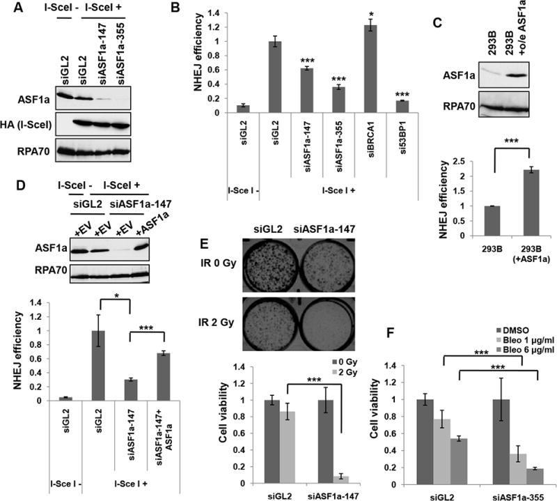
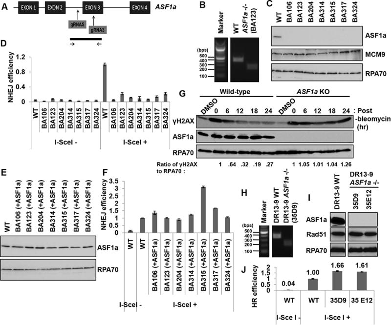
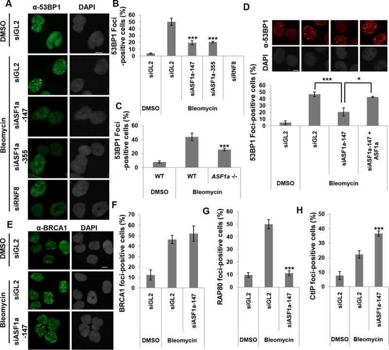
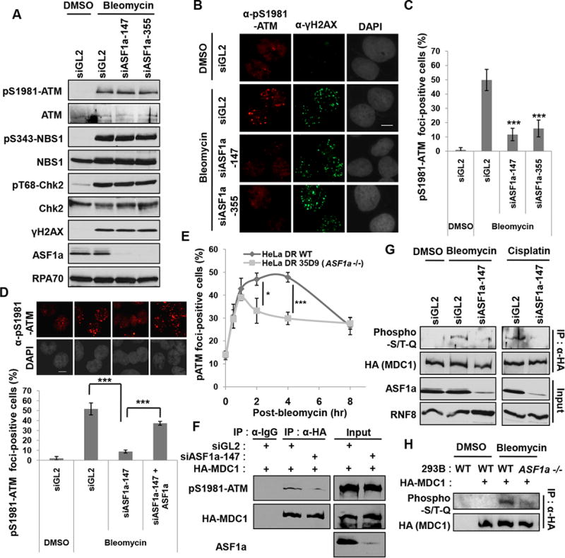
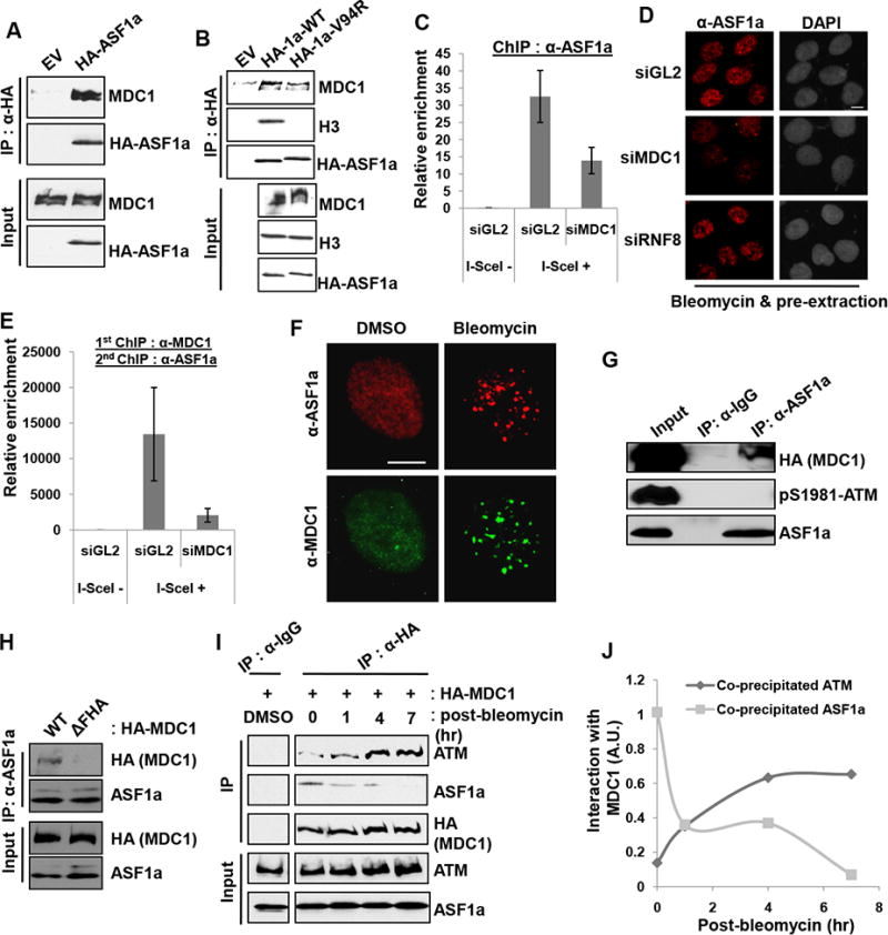
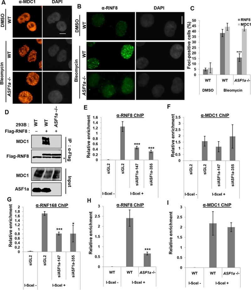
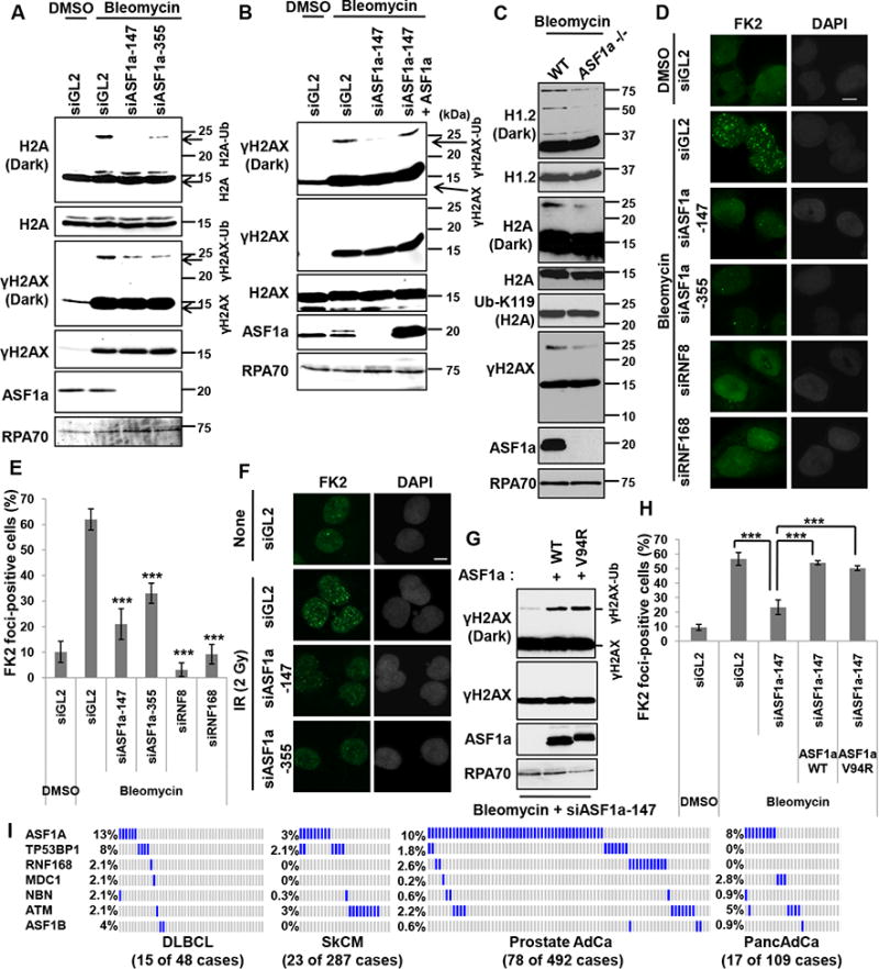
Double-strand breaks (DSBs) of DNA in eukaryotic cells are predominantly repaired by non-homologous end joining (NHEJ). The histone chaperone anti-silencing factor 1a (ASF1a) interacts with MDC1 and is recruited to sites of DSBs to facilitate the interaction of phospho-ATM with MDC1 and phosphorylation of MDC1, which are required for the recruitment of RNF8/RNF168 histone ubiquitin ligases. Thus, ASF1a deficiency reduces histone ubiquitination at DSBs, decreasing the recruitment of 53BP1, and decreases NHEJ, rendering cells more sensitive to DSBs. This role of ASF1a in DSB repair cannot be provided by the closely related ASF1b and does not require its histone chaperone activity. Homozygous deletion of ASF1A is seen in 10%-15% of certain cancers, suggesting that loss of NHEJ may be selected in some malignancies and that the deletion can be used as a molecular biomarker for cancers susceptible to radiotherapy or to DSB-inducing chemotherapy.

Keywords: 53BP1; ASF1a; ATM; HR; MDC1; NHEJ; RNF168; RNF8; histone; ubiquitination.


Figures

Figure 1
Figure 1. ASF1a is required for NHEJ and resistance to DSBs
(A) Immunoblots of the NHEJ/DsRed293B lysates transfected with two different ASF1a targeting siRNAs, 48 hr after transfection of HA-I-SceI plasmids. HA-I-SceI was detected by anti-HA antibody. (B) ASF1a knockdown reduces NHEJ efficiency. NHEJ efficiency is measured as described in the METHOD DETAILS and represented as mean ± S.D. of triplicates. ***, P< 0.005; *, P< 0.05. (C) ASF1a overexpression increases NHEJ efficiency. 293B having stable overexpression (o/e) of ASF1a was compared with wild-type 293B for ASF1a expression level in the immunoblot (top) and NHEJ efficiency (bottom). Mean ± S.D. from triplicate measurements. (D) Rescue of NHEJ in siASF1a-transfected 293B cells by expression of siRNA-resistant ASF1a. Empty (+EV) or ASF1a expressing vector resistant to siASF1a (+ASF1a) was co-transfected with HA-I-SceI. Immunoblots (top) and quantitation of NHEJ efficiency (bottom). Mean ± S.D. of triplicates. (E) Depletion of ASF1a renders cells sensitive to ionizing radiation (IR). Cell viability was quantified and presented as mean ± S.D. from triplicate measurements (lower panel). Representative images (upper panel). (F) Dose-dependent sensitivity to bleomycin of ASF1a depleted cells. The indicated dose of bleomycin was treated for 24 hr after 48 hr from first siRNA transfection. Mean ± S.D. from triplicates.
Figure 2
Figure 2. Knockout of ASF1a reduces NHEJ and promotes HR
(A) A schematic of the targeting strategy for ASF1a knockout in 293B or HeLa DR13-9 cells using the CRSPR/CAS9 system. The sgRNAs targeting the ASF1a gene (top) and the region interrogated to identify the deletion (bottom) are shown. (B) A representative image of the PCR product from the 293B clones: Wild type and BA123 (with a homozygous deletion of the ASF1a gene). (C) A western blot showing ASF1a protein level in 293B wild-type and ASF1a null clones. (D) Knocking-out ASF1a suppresses NHEJ efficiency. The percentage of DsRed-positive cells in each cell-line was normalized to that of wild-type cells transfected with HA-I-SceI. Mean ± S.D. from triplicates. (E and F) ASF1a expression in ASF1a knockout cells rescues NHEJ efficiency. ASF1a was stably expressed in ASF1a knockout cell lines using retroviral infection. Immunoblots of those lysates (E) and NHEJ assay (F). Mean ± S.D. from triplicates. (G) Decrease of γH2AX after DSB is slowed in ASF1a knockout cells. The ratio of γH2AX to RPA70 signal was quantitated at each time point and normalized to the ratio at the 0 hr point. See also Figure S1A. (H) Representative image of PCR on genomic DNA from HeLa DR13-9 clones: wild type and 35D9. (I) A western blot showing ASF1a protein level in wild-type and ASF1a null clones of HeLa DR13-9. (J) Knocking-out ASF1a promotes HR repair. HR was measured as described in the METHOD DETAILS. Mean ± S.D. from triplicates. See also Figure S1B to S1D.
Figure 3
Figure 3. ASF1a is required for the recruitment of 53BP1 and RAP80 at DSBs
(A to C) Decrease of 53BP1 foci upon depletion of ASF1a in U2OS cells or in ASF1a -/-cells after treatment with bleomycin for 1 hr before fixation. Representative images (A) and quantitation (B and C). Cells with >20 foci of 53BP1 were counted. Scale bar, 10 μm. Mean ± S.D. of triplicates. ***, P < 0.005. Scale bar, 10 μm. See also Figure S2. (D) Rescue of 53BP1 foci in ASF1a-depleted U2OS cells by siRNA-resistant ASF1a. Representative images (top) and Mean ± S.D. of triplicates. ***, P< 0.005; *, P< 0.05 (bottom). (E and F) No change of BRCA1 foci upon depletion of ASF1a. Representative images (E) and quantitation (F). (G) RAP80 foci are decreased in ASF1a depleted U2OS cells. (H) CtIP foci are increased in ASF1a depleted U2OS cells. The quantitations in (F-G) is the same as in (B), and cells having over 5 foci of CtIP were counted in (H).
Figure 4
Figure 4. ASF1a promotes the localization of ATM and phosphorylation of MDC1 at DSB by facilitating the ATM-MDC1 interaction
(A) Immunoblots with indicated antibodies show no decrease in DNA damage induced autophosphorylation of ATM or in ATM phosphorylation of NBS1, Chk2 and H2AX after depletion of ASF1a. (B and C) Decreased pS1981-ATM foci after depletion of ASF1a. Representative images of immunostaining (B) and quantification of foci-positive cells (C). Scale bar, 10 μm. Mean ± S.D. of triplicates. ***, P < 0.005. (D) siRNA-resistant ASF1a rescues pS1981-ATM foci-formation. Representative images (top) and quantification of cells >10 pS1981-ATM foci (bottom). Mean ± S.D. of triplicates. (E) A rapid disappearance of pS1981-ATM foci-formation upon ASF1a knockout. The cells having >5 pS1981-ATM foci in wild type or ASF1a -/- HeLa DR13-9 were counted as positive cells at indicated time points after pulse-treatment of 40 μgml-1 bleomycin for 20 min. Mean ± S.D. of triplicates. *, P < 0.05; ***, P < 0.005. (F) Defective interaction of MDC1 with phospho-ATM after ASF1a knockdown. (G and H) ASF1a is required for ATM phosphorylation on MDC1. HEK293T cells transfected with indicated siRNAs (G), 293B wild type and ASF1a -/- cells (H) were transfected by HA-MDC1 plasmid and DMSO, bleomycin or cisplatin was added for 14 hr before harvest.
Figure 5
Figure 5. ASF1a interacts with MDC1, and the FHA domain of MDC1 is required for ASF1a localization to DSBs
(A) HA-ASF1a precipitates contain endogenous MDC1. Immunoblots of immunoprecipitates or input lysate. (B) Histone binding by ASF1a not required for the interaction with MDC1. Wild-type or Val94Arg mutant of HA-ASF1a was immunoprecipitated with anti-HA antibody. (C) MDC1 is required for ASF1a localization at I-SceI cut site. ChIP assay was performed as described in the METHOD DETAILS. (D) MDC1 dependent association of ASF1a with chromatin after DNA damage. U2OS cells treated with bleomycin for 1 hr followed by pre-extraction, to remove soluble proteins, were fixed and immunostained. Scale bar, 10 μm. (E) Co-localization of MDC1 and ASF1a to I-SceI cut site. The eluate from anti-MDC1 ChIP was applied to an anti-ASF1a ChIP. (F) Co-localization of ASF1a- and MDC1-foci at DSBs. (G) Untagged ASF1a co-immunoprecipitates MDC1 but not phospho-ATM. (H) ASF1a-MDC1 interaction needs the FHA domain of MDC1. HA-MDC1 wild type or FHA deletion mutant (∆55-124 a.a.) was overexpressed in HEK293T cells followed by bleomycin treatment for 14 hr. (I and J) MDC1 interaction with ASF1a decreases as its interaction with ATM increases after DNA damage. The immunoblots with indicated antibodies (I) and quantification of co-precipitated ATM or ASF1a with HA-MDC1 after pulse-treatment of bleomycin (J).
Figure 6
Figure 6. ASF1a is required for RNF8, but not MDC1, recruitment at DSBs
(A to C) Decrease of bleomycin-induced RNF8 foci, but not MDC1 foci in ASF1a knockout cells. HeLa DR13-9 wild type or ASF1a -/- cells were incubated with 20 μgml-1 bleomycin for 40 min followed by fixation. Representative images of immunostaining with indicated antibodies (A and B), and quantification of cells with >5 foci of RNF8 or MDC1 (C). See colocalization of RNF8 and MDC1 foci in Figure S3. Scale bar, 10 μm. Mean ± S.D. of triplicates. ***, P < 0.005. (D) Decreased interaction of MDC1 with RNF8 after DNA damage in ASF1a knockout cells. Plasmid expressing Flag-RNF8 was transfected in 293B wild type and ASF1a -/- cells followed by bleomycin treatment before harvest. Asterisk indicates immunoglobulin heavy chain. (E to I) ASF1a is required for RNF8/168 localization, not MDC1, at I-SceI cut site. HeLa DR13-9 cells transfected with indicated siRNAs (E to G), wild type and ASF1a -/- cells (H and I) were applied to ChIP assay using anti-RNF8 (E and H), -MDC1 (F and I) and -RNF168 (G) antibodies. Mean ± SD of triplicates. ***, P < 0.005; *, P< 0.05.
Figure 7
Figure 7. DSB-dependent ubiquitination on histones and FK2 focus formation require ASF1a
(A) DSB-induced H2A or H2AX mono-ubiquitination is dependent on ASF1a. HEK293T cells were transfected with indicated siRNAs and treated with bleomycin. (B) Expression of siRNA-resistant ASF1a restores γH2AX mono-ubiquitination in cells depleted of endogenous ASF1a. Indicated siRNAs were transfected into either wild type U2OS or U2OS cells with stable overexpression of siRNA-resistant ASF1a. (C) Decrease of RNF8/168-dependent ubiquitination of H1.2, H2A and γH2AX, but not PRC1 dependent ubiquitination of H2A K119, in ASF1a knockout cells. HeLa DR13-9 wild type or ASF1a -/- cells were incubated with 20 μgml-1 bleomycin for 80 min. (D, E and F) Immunostaining with anti-FK2 antibody in ASF1a depleted cells. U2OS cells were transfected with indicated siRNA and immunostained following treatment with 5 μgml-1 bleomycin for 1 hr (D and E) or at 90 min post-IR (2 Gy) (F). Representative images (D and F) and quantification of (D) in (E). Mean ± SD of triplicates. ***, P < 0.005. (G) Rescue of mono-ubiquitination of H2AX by wild-type or Val94Arg mutant ASF1a in ASF1a-depleted U2OS cells. (H) Rescue of FK2 foci by wild-type or Val94Arg mutant ASF1a in ASF1a-depleted U2OS cells. Indicated siRNAs were transfected into either wild type U2OS or U2OS cells with stable overexpression of siRNA-resistant ASF1a wild type or V94R mutant. Cells were immunostained after treatment of 10 μgml-1 bleomycin for 1 hr. Mean ± SD of triplicates. See also Figure S5. (I) High frequency of homozygous deletion of ASF1a gene in cancers. Cbioportal (http://cbioportal.org) was analyzed and each blue or gray bar represents a patient with or without a homozygous deletion in the indicated gene, respectively. To save space a large number of gray bars were omitted from the rows in SkCM, Prostate AdCa and PancAdCa. The number of patients whose tumors had homozygous deletion of at least one of these repair genes (excluding ASF1B) is shown below in each cancer.

Similar articles

Cited by

References

    1. Battu A, Ray A, Wani AA. ASF1A and ATM regulate H3K56-mediated cell-cycle checkpoint recovery in response to UV irradiation. Nucleic Acids Res. 2011;39:7931–7945. - PMC - PubMed
    1. Bothmer A, Robbiani DF, Feldhahn N, Gazumyan A, Nussenzweig A, Nussenzweig MC. 53BP1 regulates DNA resection and the choice between classical and alternative end joining during class switch recombination. J Exp Med. 2010;207:855–865. - PMC - PubMed
    1. Botuyan MV, Lee J, Ward IM, Kim JE, Thompson JR, Chen J, Mer G. Structural basis for the methylation state-specific recognition of histone H4-K20 by 53BP1 and Crb2 in DNA repair. Cell. 2006;127:1361–1373. - PMC - PubMed
    1. Bouwman P, Aly A, Escandell JM, Pieterse M, Bartkova J, van der Gulden H, Hiddingh S, Thanasoula M, Kulkarni A, Yang Q, et al. 53BP1 loss rescues BRCA1 deficiency and is associated with triple-negative and BRCA-mutated breast cancers. Nat Struct Mol Biol. 2010;17:688–695. - PMC - PubMed
    1. Braggio E, Dogan A, Keats JJ, Chng WJ, Huang G, Matthews JM, Maurer MJ, Law ME, Bosler DS, Barrett M, et al. Genomic analysis of marginal zone and lymphoplasmacytic lymphomas identified common and disease-specific abnormalities. Mod Pathol. 2012;25:651–660. - PMC - PubMed

MeSH terms

Substances