

Diabetic Hemichorea-hemiballism after Prompt Improvement in Hyperglycemia
- PMID: 28943546
- PMCID: PMC5725863
- DOI: 10.2169/internalmedicine.8615-16
Diabetic Hemichorea-hemiballism after Prompt Improvement in Hyperglycemia
Abstract
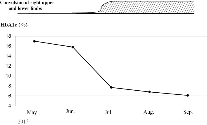
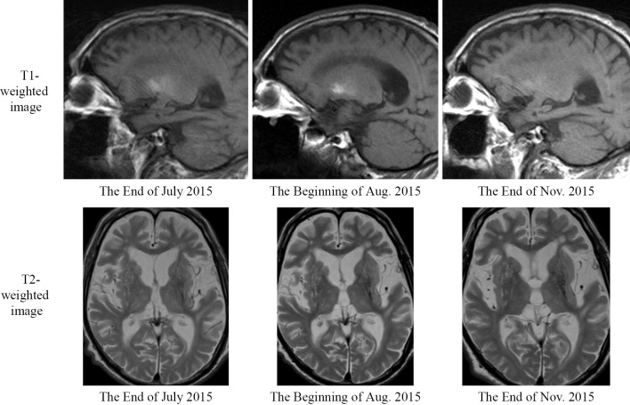
We herein report a case of hemichorea-hemiballism in an 85-year-old man diagnosed with diabetes at 76 years of age. After a one-year interruption in treatment, he was treated with a low-calorie diet, linagliptin, and nateglinide. Over 51 days, his HbA1c level decreased from 15.8% to 7.7%. After a prompt improvement in his hyperglycemia, he began experiencing involuntary movements in the right upper and lower extremities. T1-weighted magnetic resonance imaging showed a high signal intensity in the left lens nucleus. The patient was diagnosed with diabetic hemichorea-hemiballism and received haloperidol (1 mg/day) as treatment.
Keywords: diabetic hemichorea-hemiballism; lens nucleus; prompt improvement of hyperglycemia.
Figures
Similar articles
-
Diabetic striatopathy: an updated overview of current knowledge and future perspectives.J Endocrinol Invest. 2024 Jan;47(1):1-15. doi: 10.1007/s40618-023-02166-5. Epub 2023 Aug 14. J Endocrinol Invest. 2024. PMID: 37578646 Free PMC article. Review.
-
Delayed onset diabetic striatopathy: Hemichorea-hemiballism one month after a hyperglycemic episode.Am J Emerg Med. 2017 Jul;35(7):1036.e3-1036.e4. doi: 10.1016/j.ajem.2017.02.018. Epub 2017 Feb 5. Am J Emerg Med. 2017. PMID: 28202297
-
Investigation of the relationship between non-ketotic hyperglycemia and hemichorea-hemiballism: A case report.Medicine (Baltimore). 2019 Jul;98(28):e16255. doi: 10.1097/MD.0000000000016255. Medicine (Baltimore). 2019. PMID: 31305406 Free PMC article.
-
Exploration of motor cortex excitability in a diabetic patient with hemiballism-hemichorea.Mov Disord. 2000 Sep;15(5):1000-5. doi: 10.1002/1531-8257(200009)15:5<1000::aid-mds1037>3.0.co;2-e. Mov Disord. 2000. PMID: 11009213
-
[Two diabetics with hemichorea-hemiballism and striatal lesions].No To Shinkei. 1995 Feb;47(2):167-72. No To Shinkei. 1995. PMID: 7669416 Review. Japanese.
Cited by
-
Diabetic Striatopathy Complicated With Acute Ischemic Stroke: A Case Report.Front Neurosci. 2022 Jul 12;16:877479. doi: 10.3389/fnins.2022.877479. eCollection 2022. Front Neurosci. 2022. PMID: 35903807 Free PMC article.
-
Clinical features of hemichoreahemiballism: A stroke-related movement disorder.Neurol Int. 2020 Jul 10;12(1):8328. doi: 10.4081/ni.2020.8328. eCollection 2020 Jul 10. Neurol Int. 2020. PMID: 32774821 Free PMC article.
-
Relationship Between Diabetic Chorea and Timing of MRI Findings: A Systematic Review with Case Reports.Int J Gen Med. 2023 Oct 2;16:4465-4476. doi: 10.2147/IJGM.S423400. eCollection 2023. Int J Gen Med. 2023. PMID: 37808208 Free PMC article. Review.
-
A Case of Diabetic Chorea Secondary to Appetite Loss Due to COVID-19 Vaccination.Cureus. 2023 Nov 20;15(11):e49138. doi: 10.7759/cureus.49138. eCollection 2023 Nov. Cureus. 2023. PMID: 38130532 Free PMC article.
-
Diabetic striatopathy: an updated overview of current knowledge and future perspectives.J Endocrinol Invest. 2024 Jan;47(1):1-15. doi: 10.1007/s40618-023-02166-5. Epub 2023 Aug 14. J Endocrinol Invest. 2024. PMID: 37578646 Free PMC article. Review.
References
-
- Nakajima N, Ueda M, Nagayama H, Katayama Y. Putaminal changes before the oncet of clinical symptoms in diabetic hemichorea-hemiballism. Intern Med 53: 489-491, 2014. - PubMed
-
- Iwata A, Koike F, Arasaki K, Tamaki M. Blood brain barrier destruction in hyperglycemic chorea in a patient with poorly controlled diabetes. J Neurol Sci 163: 90-93, 1999. - PubMed
Publication types
MeSH terms
Substances
LinkOut - more resources
Full Text Sources
Other Literature Sources
Medical