

Dysbiosis of the Urinary Microbiota Associated With Urine Levels of Proinflammatory Chemokine Interleukin-8 in Female Type 2 Diabetic Patients
- PMID: 28943876
- PMCID: PMC5603796
- DOI: 10.3389/fimmu.2017.01032
Dysbiosis of the Urinary Microbiota Associated With Urine Levels of Proinflammatory Chemokine Interleukin-8 in Female Type 2 Diabetic Patients
Abstract
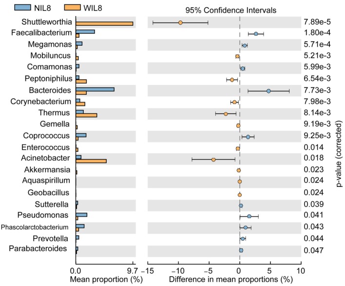
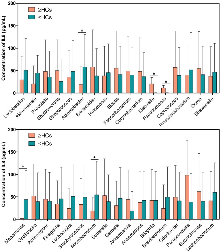
Evidence has shown that dysbiosis of the urinary microbiota existed in female type 2 diabetes mellitus (T2DM) patients. Perturbations of intestinal microbiota are linked to proinflammatory chemokine interleukin-8 (IL-8); however, the correlations between urinary microbiota and IL-8 are not well studied. Here, we investigated the associations between the altered urinary microbiota and urinary IL-8 in female T2DM patients. A modified four-tube midstream urine technique was used to collect urine specimens from 70 female T2DM patients and 70 matched healthy controls (HCs). Bacterial genomic DNA from urine specimens was isolated using magnetic beads and the urinary microbiota was assessed using Illumina MiSeq platform targeting on the 16S rRNA gene V3-V4 region. Urinary IL-8 was determined by enzyme linked immunosorbent assay. Subsequently, the T2DM patients were separated into urine IL-8 detectable (WIL8) and undetectable (NIL8) groups, and the composition of urinary microbiota between the two groups was compared. Meanwhile, the levels of IL-8 between the "≥HCs" group (those specific bacterial genera were more than or equal to the HCs) and the "<HCs" group (those specific bacterial genera were less than the HCs) was also compared. Of 70 urine samples from T2DM patients without urinary tract infections, 46 patients had detectable IL-8 in their urine (64.31 ± 70.43 pg/mL), while 24 patients had undetectable IL-8. Compared to the NIL8 group, 11 bacterial genera increased in the WIL8 group, including Corynebacterium, Akkermansia, Enterococcus, etc., whereas 10 genera, such as Faecalibacterium, Bacteroides, and Pseudomonas decreased. One species of Lactobacillus, Lactobacillus iners, increased obviously in the WIL8 group. The "≥HCs" group showed 17 genera increased and 16 genera decreased. In addition, 18 genera contributed to the presence of urinary IL-8 in T2DM patients, which explained 95.60% of the total variance of urinary microbiota. Our study demonstrated that dysbiosis of the urinary microbiota with several key bacteria was associated with urinary IL-8 in female T2DM patients, which might be useful to explore the interactions between urinary microbiota and inflammatory responses and shed light on novel diagnosis and therapy for urinary microbiota associated with infections in T2DM patients.
Keywords: Akkermansia; Lactobacillus; interleukin-8; type 2 diabetes mellitus; urinary microbiota.
Figures
Similar articles
-
Dysbiosis of urinary microbiota is positively correlated with type 2 diabetes mellitus.Oncotarget. 2017 Jan 17;8(3):3798-3810. doi: 10.18632/oncotarget.14028. Oncotarget. 2017. PMID: 28008148 Free PMC article.
-
Moderation effects of food intake on the relationship between urinary microbiota and urinary interleukin-8 in female type 2 diabetic patients.PeerJ. 2020 Jan 28;8:e8481. doi: 10.7717/peerj.8481. eCollection 2020. PeerJ. 2020. PMID: 32025384 Free PMC article.
-
Alterations of Urinary Microbiota in Type 2 Diabetes Mellitus with Hypertension and/or Hyperlipidemia.Front Physiol. 2017 Mar 3;8:126. doi: 10.3389/fphys.2017.00126. eCollection 2017. Front Physiol. 2017. PMID: 28316574 Free PMC article.
-
The human urinary microbiome and how it relates to urogynecology.Int Urogynecol J. 2016 Sep;27(9):1307-12. doi: 10.1007/s00192-016-2944-5. Epub 2016 Jan 25. Int Urogynecol J. 2016. PMID: 26811114 Review.
-
The Role of Beneficial Microbiota in COVID-19: Insights from Key Bacterial Genera.Microorganisms. 2025 Apr 29;13(5):1029. doi: 10.3390/microorganisms13051029. Microorganisms. 2025. PMID: 40431202 Free PMC article. Review.
Cited by
-
Native and Engineered Probiotics: Promising Agents against Related Systemic and Intestinal Diseases.Int J Mol Sci. 2022 Jan 6;23(2):594. doi: 10.3390/ijms23020594. Int J Mol Sci. 2022. PMID: 35054790 Free PMC article. Review.
-
Urinary microbiome profiling as a non-invasive tool for identifying biomarkers in systemic lupus erythematosus and lupus nephritis.Front Cell Infect Microbiol. 2024 Dec 3;14:1364333. doi: 10.3389/fcimb.2024.1364333. eCollection 2024. Front Cell Infect Microbiol. 2024. PMID: 39691697 Free PMC article.
-
Dysbiosis of the Human Urinary Microbiome and its Association to Diseases Affecting the Urinary System.Indian J Microbiol. 2022 Jun;62(2):153-166. doi: 10.1007/s12088-021-00991-x. Epub 2021 Nov 16. Indian J Microbiol. 2022. PMID: 35462710 Free PMC article. Review.
-
A cross-sectional study on the effects of intravesical BCG on urinary microbiota in bladder cancer patients.Int Urol Nephrol. 2025 Jun 30. doi: 10.1007/s11255-025-04607-x. Online ahead of print. Int Urol Nephrol. 2025. PMID: 40588703
-
Multiple bacteria associated with the more dysbiotic genitourinary microbiomes in patients with type 2 diabetes mellitus.Sci Rep. 2021 Jan 19;11(1):1824. doi: 10.1038/s41598-021-81507-x. Sci Rep. 2021. PMID: 33469094 Free PMC article.
References
-
- World Health Organization. Global Report on Diabetes. Geneva, Switzerland: WHO Press; (2016).
LinkOut - more resources
Full Text Sources
Other Literature Sources