

Current and Future Methods for Measuring Breast Density: A Brief Comparative Review
- PMID: 28943893
- PMCID: PMC5609705
- DOI: 10.2217/bmt.15.13
Current and Future Methods for Measuring Breast Density: A Brief Comparative Review
Abstract
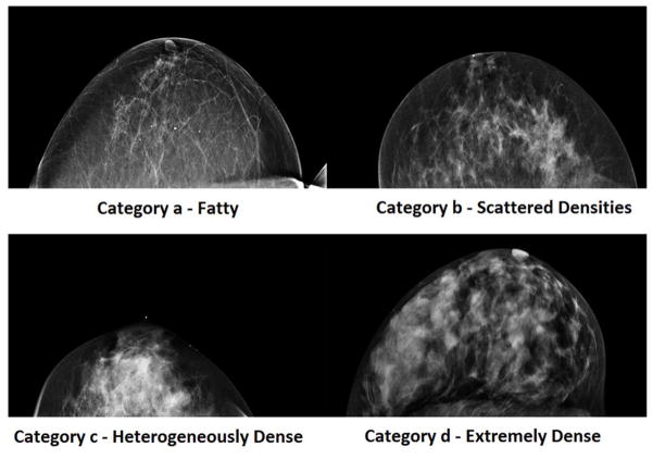
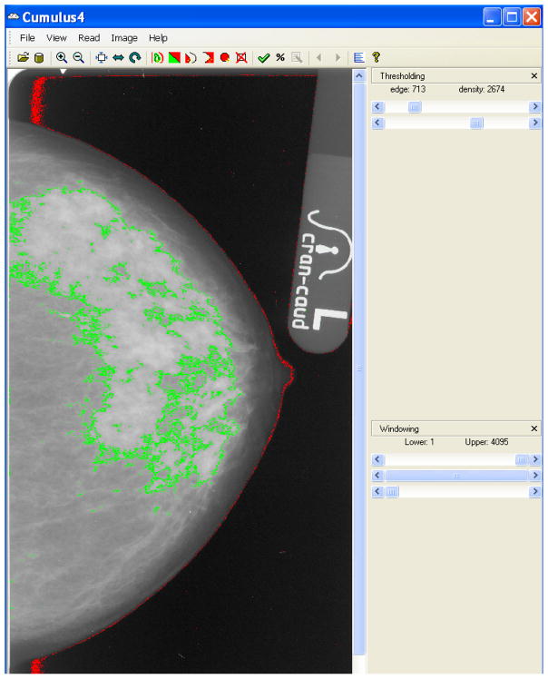
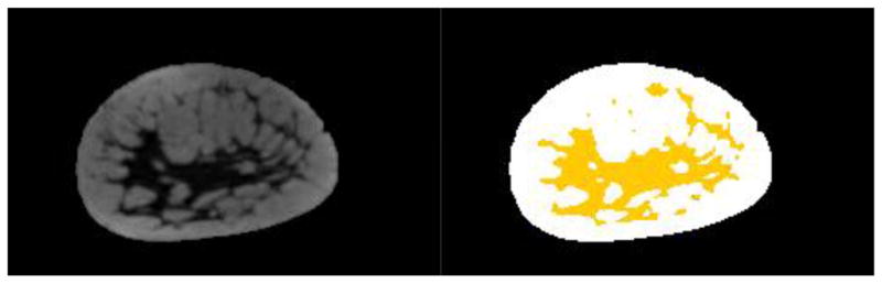
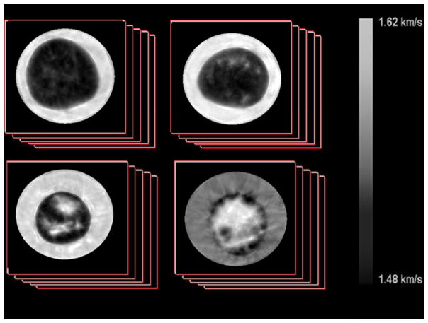
Breast density is one of the strongest predictors of breast cancer risk. Women with the densest breasts are 4 to 6 times more likely to develop cancer compared with those with the lowest densities. Breast density is generally assessed using mammographic imaging; however, this approach has limitations. Magnetic resonance imaging and ultrasound tomography are some alternative imaging modalities that can aid mammography in patient screening and the measurement of breast density. As breast density becomes more commonly discussed, knowledge of the advantages and limitations of breast density as a marker of risk will become more critical. This review article discusses the relationship between breast density and breast cancer risk, lists the benefits and drawbacks of using multiple different imaging modalities to measure density and briefly discusses how breast density will be applied to aid in breast cancer prevention and treatment.
Keywords: Breast cancer risk; Breast density; Mammography; Ultrasound tomography.
Figures
References
-
- Berry DA, Cronin KA, Plevritis SK, et al. Effect of Screening and Adjuvant Therapy on Mortality from Breast Cancer. New England Journal of Medicine. 2005;353(17):1784–1792. - PubMed
-
- Gail MH, Brinton LA, Byar DP, et al. Projecting Individualized Probabilities of Developing Breast Cancer for White Females Who Are Being Examined Annually. Journal of the National Cancer Institute. 1989;81(24):1879–1886. - PubMed
-
- American Cancer Society. Breast cancer facts and figures 2013–2014. Atlanta: American Cancer Society, Inc; 2013.
Grants and funding
LinkOut - more resources
Full Text Sources
Other Literature Sources
Research Materials