

Influence of chronic inflammation on Bcl-2 and PCNA expression in prostate needle biopsy specimens
- PMID: 28943900
- PMCID: PMC5604163
- DOI: 10.3892/ol.2017.6668
Influence of chronic inflammation on Bcl-2 and PCNA expression in prostate needle biopsy specimens
Abstract
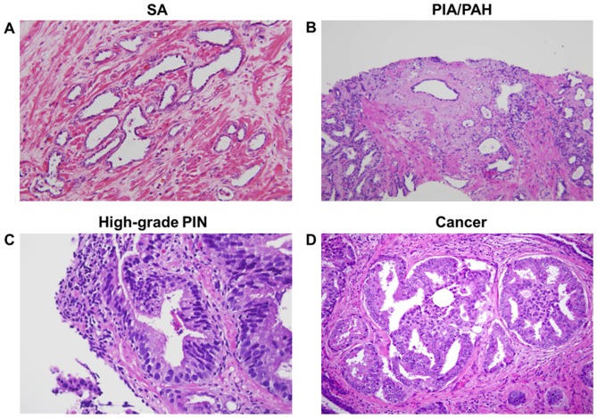
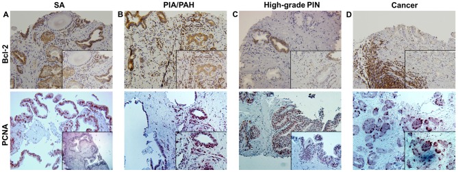
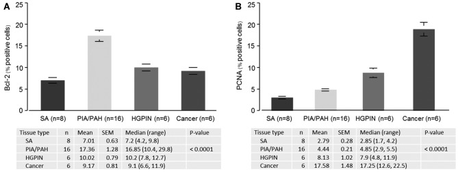
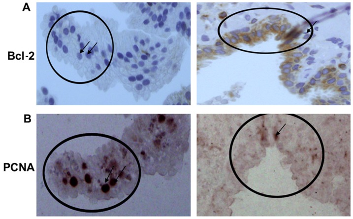
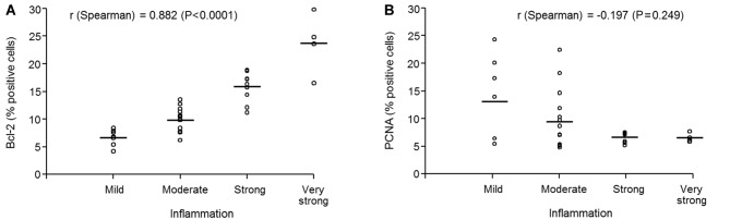
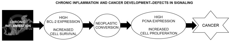
The association between inflammation and cancer has been established in certain forms of human malignancies; however, its role in prostate cancer remains unclear. The present study investigates a possible association between chronic inflammation and the development of epithelial neoplasia in the prostate. Needle biopsy specimens were obtained from patients with serum prostate-specific antigen levels >4 ng/ml, evaluated for morphological findings, and immunostained for Bcl-2 and proliferating cell nuclear antigen (PCNA). Bcl-2 is a survival protein that appears to lie at a nodal point in pathways involved in cell survival, carcinogenesis, and development of therapeutic resistance in certain cancer types. Similarly, PCNA is a critical protein for DNA replication, repair of DNA damage, chromatin structure maintenance, chromosome segregation and cell-cycle progression. The association between these two proteins was examined in prostate tissues with and without chronic inflammation, as well as tissues with and without evidence of neoplastic changes. Of the 106 needle biopsies examined, 18% exhibited atrophy with inflammation. Proliferative inflammatory atrophy/post-atrophic hyperplasia were observed in 42%, high-grade prostatic intraepithelial neoplasia (HGPIN) in 8%, prostatic adenocarcinoma in 11%, and 2% had atypical acinar proliferation suspicious for malignancy. A total of 36 specimens were stained for Bcl-2 and PCNA. Bcl-2 was expressed widely in inflammatory and epithelial tissue; however, more intense expression was observed in the areas of chronic inflammation, predominantly in infiltrating immune cells. The highest proliferation index was observed in the epithelia of HGPIN and cancer. An inverse correlation between the expression of Bcl-2 and the expression of PCNA was observed in the epithelium. The areas of chronic inflammation were associated with increased Bcl-2 expression, whereas the highly proliferative epithelium minimally expressed Bcl-2. These results suggest that Bcl-2 alters the phenotype of particular epithelial cells with a gain in neoplastic characteristics, leading to a likely precursor that may later progress into HGPIN and cancer.
Keywords: chronic inflammation; needle biopsy; post atrophic hyperplasia; proliferative inflammatory atrophy; prostate cancer; prostate-specific antigen.
Figures
Similar articles
-
Correlation of increased apoptosis and proliferation with development of prostatic intraepithelial neoplasia (PIN) in ventral prostate of the Noble rat.Prostate. 2000 Jun 15;44(1):31-9. doi: 10.1002/1097-0045(20000615)44:1<31::aid-pros5>3.0.co;2-o. Prostate. 2000. PMID: 10861755
-
The influence of chronic inflammation in prostatic carcinogenesis: a 5-year followup study.J Urol. 2006 Sep;176(3):1012-6. doi: 10.1016/j.juro.2006.04.033. J Urol. 2006. PMID: 16890681
-
Morphological transition of proliferative inflammatory atrophy to high-grade intraepithelial neoplasia and cancer in human prostate.Prostate. 2009 Sep 15;69(13):1378-86. doi: 10.1002/pros.20992. Prostate. 2009. PMID: 19507201
-
Prostate needle biopsies containing prostatic intraepithelial neoplasia or atypical foci suspicious for carcinoma: implications for patient care.J Urol. 2006 Mar;175(3 Pt 1):820-34. doi: 10.1016/S0022-5347(05)00337-X. J Urol. 2006. PMID: 16469560 Review.
-
Histological markers of risk and the role of high-grade prostatic intraepithelial neoplasia.Urology. 2001 Apr;57(4 Suppl 1):115-20. doi: 10.1016/s0090-4295(00)00953-5. Urology. 2001. PMID: 11295607 Review.
Cited by
-
Impact of e-cigarettes on colonic mucosa and the role of recovery: involvement of oxidative and inflammatory pathway.Environ Sci Pollut Res Int. 2021 Dec;28(45):64561-64571. doi: 10.1007/s11356-021-15575-x. Epub 2021 Jul 26. Environ Sci Pollut Res Int. 2021. PMID: 34312757 Free PMC article.
-
Korean Red Ginseng and Korean black ginseng extracts, JP5 and BG1, prevent hepatic oxidative stress and inflammation induced by environmental heat stress.J Ginseng Res. 2020 Mar;44(2):267-273. doi: 10.1016/j.jgr.2018.12.005. Epub 2018 Dec 16. J Ginseng Res. 2020. PMID: 32148408 Free PMC article.
-
Eriochloa villosa Alleviates Progression of Benign Prostatic Hyperplasia in vitro and in vivo.Res Rep Urol. 2022 Sep 24;14:313-326. doi: 10.2147/RRU.S381713. eCollection 2022. Res Rep Urol. 2022. PMID: 36187165 Free PMC article.
-
High expression of ladinin-1 (LAD1) predicts adverse outcomes: a new candidate docetaxel resistance gene for prostatic cancer (PCa).Bioengineered. 2021 Dec;12(1):5749-5759. doi: 10.1080/21655979.2021.1968647. Bioengineered. 2021. PMID: 34516317 Free PMC article.
-
Histoprotective Effect of Essential Oil from Citrus aurantifolia in Testosterone-Induced Benign Prostatic Hyperplasia Rat.Adv Urol. 2019 Sep 25;2019:3031609. doi: 10.1155/2019/3031609. eCollection 2019. Adv Urol. 2019. PMID: 31662741 Free PMC article.
References
-
- Umbehr MH, Gurel B, Murtola TJ, Sutcliffe S, Peskoe SB, Tangen CM, Goodman PJ, Thompson IM, Lippman SM, Lucia MS, et al. Intraprostatic inflammation is positively associated with serum PSA in men with PSA <4 ng ml(−1), normal DRE and negative for prostate cancer. Prostate Cancer Prostatic Dis. 2015;18:264–269. doi: 10.1038/pcan.2015.19. - DOI - PMC - PubMed
Grants and funding
LinkOut - more resources
Full Text Sources
Other Literature Sources
Miscellaneous