. 2017 Oct;14(4):4183-4189.
doi: 10.3892/ol.2017.6719. Epub 2017 Aug 4.

Combination of galectin-3, CK19 and HBME-1 immunostaining improves the diagnosis of thyroid cancer

Affiliations

Combination of galectin-3, CK19 and HBME-1 immunostaining improves the diagnosis of thyroid cancer

Vanessa Arcolia et al. Oncol Lett. 2017 Oct.

Abstract

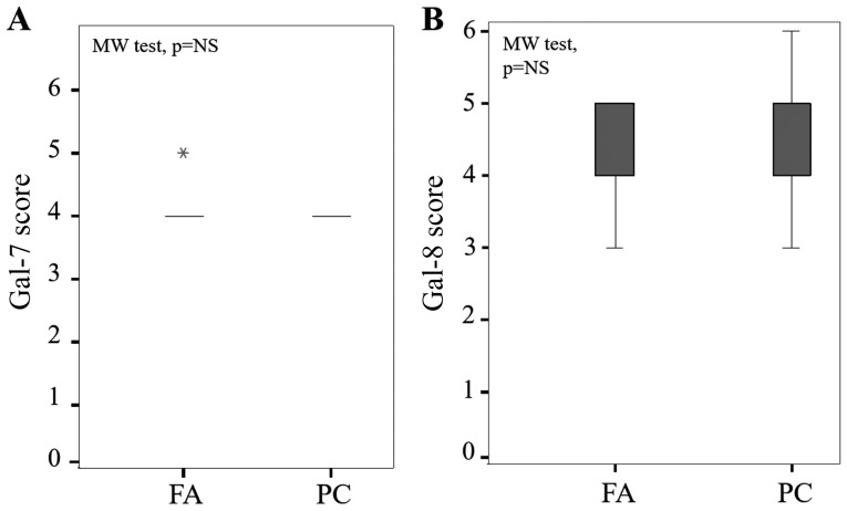
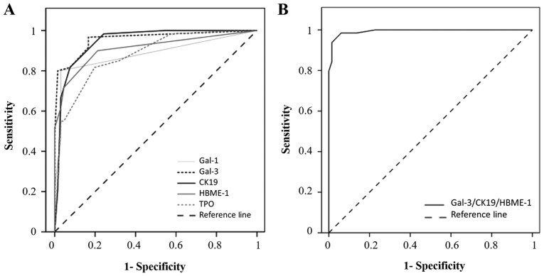
Currently, fine-needle aspiration is the most frequently used pre-operative technique for diagnosis of malignant thyroid tumors, however, pathologists are unable to reach efficient and accurate differential diagnoses between benign and malignant thyroid nodules. To aid in resolving this issue, immunohistochemistry for galectins (gal)-1, -3, -7, -8, cytokeratin 19 (CK19), Hector Battifora Mesothelial Epitope-1 (HBME-1) and thyroid peroxidase (TPO) was performed on two tissue microarrays composed of 66 follicular adenomas (FA) and 66 papillary carcinomas (PC). The identification of optimal cut-off levels and the diagnostic value of single immunomarkers or combinations were evaluated using the receiver operating characteristic curve analysis. Signal intensities for gal-1, gal-3, CK19 and HBME-1 were significantly greater in PC compared with FA (P<0.001). Conversely, expression levels of TPO were significantly increased in FA compared with PC (P<0.001). Gal-3 and CK19 appeared to be the most sensitive markers (97 and 98%, respectively), whereas galectin-1 was the most specific (97%). The combination of gal-3, CK19 and HBME-1 acted as the most efficient and informative marker panel reaching the greatest specificity (97%) and sensitivity (95%) for the diagnosis of PCs. The findings suggest that this combination of markers may improve the reliability of diagnosis of thyroid cancer.

Keywords: ROC curve; cytokeratin; galectin; lectin; malignancy.


Figures

Figure 1.
Figure 1.
Evaluation of gal-3, CK19, HBME-1, gal-1 and TPO expression in benign and malignant thyroid lesions. Immunohistochemical evaluation of expression and statistical analysis of (A) gal-3, (B) CK19, (C) HBME-1, (D) gal-1 and (E) TPO in tissue microarray composed by FA and PC (P<0.001, Mann-Whitney test). Magnification, ×400. The immunostaining was semi-quantitatively assessed in the cytoplasmic compartment and scored from 0 to 6 by summing staining intensity (0–3) and percentage of positivity (0–3). Data are presented as box plots indicating the 1st and the 3rd quartiles centered on medians (thick lines) with whiskers for the minimum and maximum non-outlier values, the ‘o’ symbols are outliers and the ‘*’ symbols show the extreme values. Gal-3, galectin-3; CK19, cytokeratin 19; HBME-1, Hector Battifora Mesothelial Epitope-1; TPO, thyroid peroxidase; FA, follicular adenoma; PC, papillary carcinoma.
Figure 2.
Figure 2.
Evaluation of gal-7 and gal-8 expression in FA and PC. Statistical analysis of (A) gal-7 and (B) gal-8 in a series of 66 cases of FA and 66 cases of PCs (P=NS, Mann-Whitney test). The immunostaining was semi-quantitatively assessed in the cytoplasmic compartment and scored from 0 to 6 by summing staining intensity (0–3) and percentage of positivity (0–3). Data are presented as box plots indicating the 1st and the 3rd quartiles centered on medians (thick lines) with whiskers for the minimum and maximum non-outlier values, the ‘*’ symbol shows the extreme values. Gal-7, galectin-7; FA, follicular adenoma; PC, papillary carcinoma.
Figure 3.
Figure 3.
Receiver operating characteristic curves of single and combined thyroid markers. (A) ROC curve for positive immunocytochemistry expression of gal-1, gal-3, CK19, HBME-1 and TPO in the diagnosis of PCs. Areas under the curve: Gal-3=0.957; CK19=0.047; HBME-1=0.910; gal-1= 0.883; TPO=0.879. (B) ROC curve for positive immunohistochemistry expression of the combination of three markers (gal-3/CK19/HBME-1) in the diagnosis of PCs. Area under the curve, 0.994. ROC, receiver operating characteristic; gal-1, galectin-1; CK19, cytokeratin 19; HBME-1, Hector Battifora Mesothelial Epitope-1; TPO, thyroid peroxidase; PC, papillary carcinoma.

Similar articles

Cited by

References

    1. Hegedüs L. Thyroid ultrasonography as a screening tool for thyroid disease. Thyroid. 2004;14:879–880. doi: 10.1089/thy.2004.14.879. - DOI - PubMed
    1. Gharib H, Papini E. Thyroid nodules: Clinical importance, assessment and treatment. Endocrinol Metab Clin North Am. 2007;36(vi):707–735. doi: 10.1016/j.ecl.2007.04.009. - DOI - PubMed
    1. Williams ED. Guest editorial: Two proposals regarding the terminology of thyroid tumors. Int J Surg Pathol. 2000;8:181–183. doi: 10.1177/106689690000800304. - DOI - PubMed
    1. Liu Z, Li X, Shi L, Maimaiti Y, Chen T, Li Z, Wang S, Xiong Y, Guo H, He W, et al. Cytokeratin 19, thyroperoxidase, HBME-1 and galectin-3 in evaluation of aggressive behavior of papillary thyroid carcinoma. Int J Clin Exp Med. 2014;7:2304–2308. - PMC - PubMed
    1. Flanagan JN, Pineda P, Knapp PE, De Las Morenas A, Lee SL, Braverman LE. Expression of cytokeratin 19 in the diagnosis of thyroid papillary carcinoma by quantitative polymerase chain reaction. Endocr Pract. 2008;14:168–174. doi: 10.4158/EP.14.2.168. - DOI - PubMed