. 2017 Feb 21:5:16056.
doi: 10.1038/boneres.2016.56. eCollection 2017.

Exercise-induced irisin in bone and systemic irisin administration reveal new regulatory mechanisms of bone metabolism

Affiliations

Exercise-induced irisin in bone and systemic irisin administration reveal new regulatory mechanisms of bone metabolism

Jin Zhang et al. Bone Res. .

Abstract

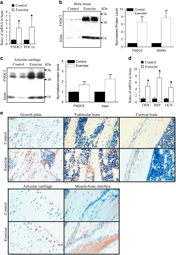
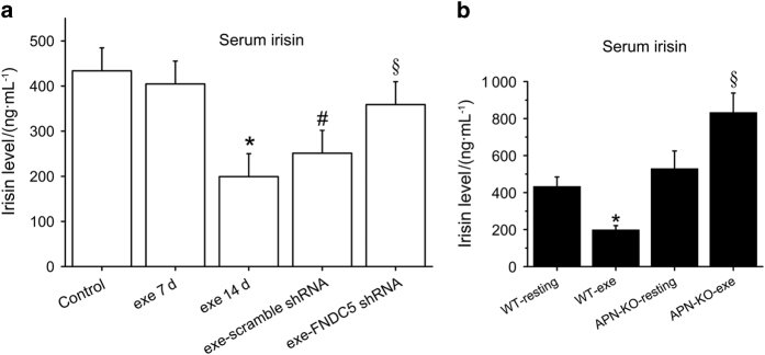
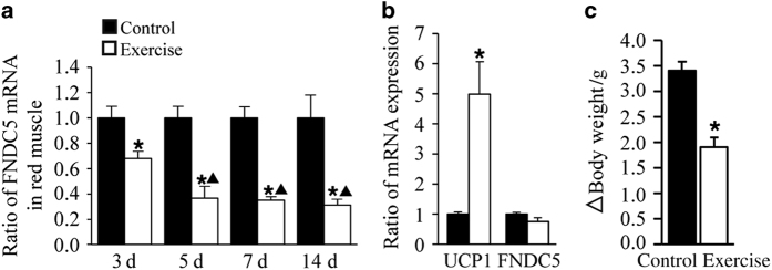
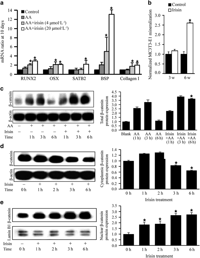
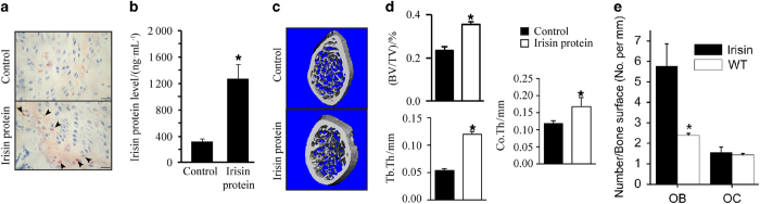
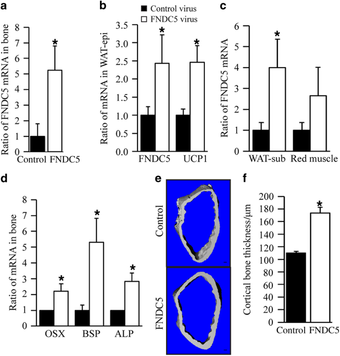
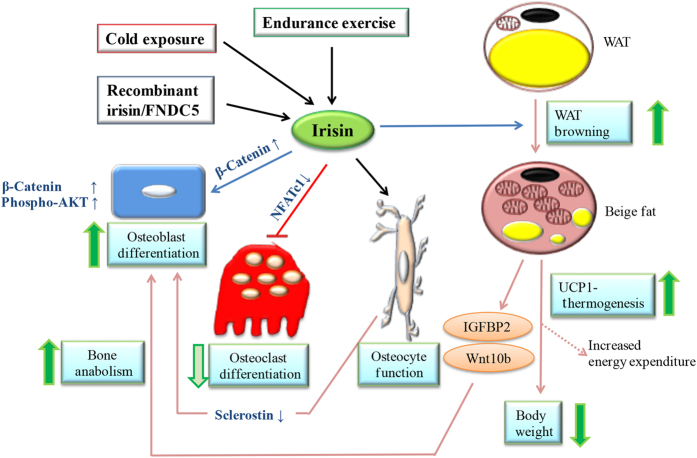
Irisin is a polypeptide hormone derived from the proteolytic cleavage of fibronectin-type III domain-containing 5 (FNDC5) protein. Once released to circulation upon exercise or cold exposure, irisin stimulates browning of white adipose tissue (WAT) and uncoupling protein 1 (UCP1) expression, leading to an increase in total body energy expenditure by augmented UCP1-mediated thermogenesis. It is currently unknown whether irisin is secreted by bone upon exercise or whether it regulates bone metabolism in vivo. In this study, we found that 2 weeks of voluntary wheel-running exercise induced high levels of FNDC5 messenger RNA as well as FNDC5/irisin protein expression in murine bone tissues. Increased immunoreactivity due to exercise-induced FNDC5/irisin expression was detected in different regions of exercised femoral bones, including growth plate, trabecular bone, cortical bone, articular cartilage, and bone-tendon interface. Exercise also increased expression of osteogenic markers in bone and that of UCP1 in WAT, and led to bodyweight loss. Irisin intraperitoneal (IP) administration resulted in increased trabecular and cortical bone thickness and osteoblasts numbers, and concurrently induced UCP1 expression in subcutaneous WAT. Lentiviral FNDC5 IP administration increased cortical bone thickness. In vitro studies in bone cells revealed irisin increases osteoblastogenesis and mineralization, and inhibits receptor activator of nuclear factor-kB ligand (RANKL)-induced osteoclastogenesis. Taken together, our findings show that voluntary exercise increases irisin production in bone, and that an increase in circulating irisin levels enhances osteogenesis in mice.


Conflict of interest statement

The authors declare no conflict of interest.

Figures

Figure 1
Figure 1
Voluntary exercise for 2 weeks increased FNDC5, PGC1α, and irisin expression in bone. Five-week-old male wild-type C57BL/6J mice weighing 17–20 g were randomly housed individually in empty cages (control group, n=18 mice) or in cages with a polycarbonate running wheel for voluntary running (exercise group, n=18 mice) for up to 2 weeks. Bone marrow was removed from bone tissues before qRT-PCR and western blot analysis. (a) qRT-PCR analysis of FNDC5 and PGC1-α mRNA expression in exercised and control bone tissue (*P<0.05, vs control). (b, c) Western blot analysis of FNDC5 and irisin protein expression in (b) exercised and control bone tissue and in (c) exercised and control articular cartilage (**P<0.01, vs control). (d) qRT-PCR analysis of osteogenic markers osterix (OSX), BSP, and osteocalcin (OCN) mRNA in bones of control and exercise group. (*P<0.05, vs control). (e) Representative immunohistochemistry with anti-irisin antibody reveals immunostaining in growth plate, trabecular bone, cortical bone, articular cartilage, and bone–muscle interface of control and exercised bones.
Figure 2
Figure 2
Voluntary exercise for 2 weeks increased serum irisin levels in mice lacking adiponectin expression, whereas decreased them in wild-type mice. Five-week-old male wild-type C57BL/6J mice weighing 17–20 g were randomly housed individually in empty cages (control group, n=18 mice) or in cages with a polycarbonate running wheel for voluntary running for 1 week (exe7d group, n=5 mice) and 2 weeks (exe14d group, n=18 mice). Five-week-old wild-type mice were injected IP with FNDC5 shRNA (exe-FNDC5 shRNA group, n=5 mice) or scrambled shRNA (exe-scrambled shRNA, n=5 mice) and then exercised for 2 weeks. (a) Serum levels of irisin were evaluated by ELISA in wild-type mice subjected to exercise for 7 and 14 days or subjected to FNDC5 shRNA or scramble shRNA treatments and 2 weeks of exercise. (*P<0.05, vs control; #P<0.05 vs control; §P<0.05 vs exe14d group). (b) Five-week-old male knockout mice (APN-KO) and wild-type mice (WT) were subjected to 2 weeks of voluntary wheel running (n=5 mice for each resting group and n=5 mice for each of the exercised groups). Serum irisin levels were evaluated and compared by ELISA between wild-type and and APN-KO groups. (*P<0.05, vs control; §P<0.05 vs APN-KO-resting).
Figure 3
Figure 3
Voluntary exercise decreased FNDC5 mRNA expression in muscle, increased UCP1 mRNA in subcutaneous WAT (WAT-sub), and decreased bodyweight in mice after 2 weeks. (a) qRT-PCR analysis of FNDC5 expression in red muscle collected at 3, 5, 7, and 14 days from exercise and control groups. (*P<0.05 vs control; ▲P<0.05 vs 3 days). (b) qRT-PCR analysis of UCP1 and FNDC5 expression in WAT-sub at day 14 from exercise and control groups. (*P<0.05 vs control). (c) Exercise and control group body weight was measured on day 0 and day 14. Changes in body weight after 2 weeks are reported (*P<0.05 vs control). All values are expressed as means±s.d. (n=18 mice per group).
Figure 4
Figure 4
Irisin increased osteoblast differentiation and nuclear levels of β-catenin in MC3T3-E1 preosteoblastic cells. (a) Irisin increased ascorbic acid (AA)-induced osteoblast differentiation genes Runt-related transcription factor 2 (RUNX2), osterix (OSX), and special AT-rich sequence-binding protein 2 (SATB2) expression, and extracellular matrix proteins bone sialoprotein (BSP) and collagen I at 10 days. (*P<0.05, vs control). (b) Mineralization assay of MC3T3-E1 cells cultured in the presence and absence of irisin for 3 and 6 weeks (*P<0.05, vs control). (c) Western blot analysis to evaluate total β-catenin protein expression in MC3T3-E1 treated in the presence of AA for 1, 3, and 6 h with or without irisin and in the absence of AA or irisin (first lane). β-Actin was used as a loading control. (d) Western blot analysis to evaluate β-catenin protein expression in cytoplasmic extracts of MC3T3-E1 cultures treated with irisin for 0, 1, 2, 3, and 6 h. β-Actin was used as a loading control. (e) Western blot analysis to evaluate β-catenin protein expression in nuclear extracts of MC3T3-E1 cultures. Lamin B1 was used as a loading control. All values are expressed as means±s.d. (*P<0.05 vs control).
Figure 5
Figure 5
Irisin reduced RANKL-induced osteoclastogenesis by inhibiting nuclear factor of activated T cells c1 (NFATc1) expression in RANKL-treated RAW264.7 cells. (a) qRT-PCR analysis of osteoclast differentiation markers cathepsin K and tartrate-resistant acid phosphatase (TRAP) in RAW264.7 cells treated with RANKL and/or irisin for 3 days (*P<0.05, vs RANKL). (b) Irisin effects on formation of multinucleated TRAP+ cells in RAW264.7 cells untreated (blank), or treated with RANKL in the presence and absence of different concentrations of irisin for 6 days. Only TRAP-positive cells with three or more nuclei were manually counted and included in the analysis. (c) Irisin effects in NFATc1 mRNA expression and NFATc1 protein expression in RAW264.7 cells untreated (blank), or treated with RANKL in the presence and absence of different concentrations of irisin for 6 days. (*P<0.05, **P<0.01, vs RANKL). (d) Western blot image and quantification of calcineurin and p-AKT1 in RANKL-induced RAW264.7 cells treated with or without irisin for 0, 10, 30, and 60 min. β-Actin was used as a loading control. All values are expressed as means±s.d. (*P<0.05, vs RANKL).
Figure 6
Figure 6
IP injection of irisin increases trabecular bone mass and osteoblast numbers in mice. (a) Representative immunohistochemistry with anti-FNDC5/irisin antibody illustrates positive cells in bones of control and irisin-treated mice for 14 days. FNDC5/Irisin-positive osteoblasts (arrows) were found on the edge of growth plate in irisin-treated mice. (b) Circulatory levels of irisin were evaluated in control and irisin-treated mice by ELISA. (*P<0.05, vs control). (c) Representative μCT images of the distal metaphyseal regions of femora of control and irisin-treated mice (n=6). Scale bars, 100 μm. (d) Trabecular bone volume/total volume (BV/TV), trabecular thickness (Tb.Th), and cortical thickness (Co.Th) were measured by μCT in femurs of control and irisin-treated mice. Values are means±s.d. of six mice per group (*P<0.05, vs control). (e) Evaluation of osteoblast (OB) and osteoclast numbers (OC) in control and irisin-treated mice. Values are means±s.d. of six mice per group (*P<0.05, vs control).
Figure 7
Figure 7
FNDC5 overexpression by IP injection of FNDC5 lentiviral particles induces expression of osteogenic markers and FNDC5 mRNA and increases cortical bone thickness. (a) qRT-PCR analysis of FNDC5 mRNA expression in bones of FNDC5 virus and control virus groups. (b) qRT-PCR analysis of FNDC5 and UCP1 mRNA expression in epididymal WAT. (c) qRT-PCR analysis of FNDC5 mRNA expression in subcutaneous WAT and red muscle. (d) qRT-PCR analysis of osteogenic markers osterix (OSX), bone sialoprotein (BSP), and ALP mRNA expression in bones (*P<0.05, vs control). (e) Representative μCT images of the distal metaphyseal regions of femora of control and irisin-treated mice (n=5). Scale bars, 100 μm. (f) Cortical thickness was measured by μCT in femurs of FNDC5 virus and control virus groups. Values are means±s.d. of five mice per group (*P<0.05, vs control).
Figure 8
Figure 8
Proposed model of irisin direct and indirect effects on bone metabolism. Exercise, cold exposure, administration with recombinant irisin, or overexpressing FNDC5 can potentially lead to increased levels of irisin in circulation according to the bibliography. In our study, 2 weeks of voluntary exercise increased expression of FNDC5/Irisin and osteogenic markers in bone (Figure 1), increased serum irisin levels in mice lacking adiponectin expression (Figure 2), and upregulated UCP1 expression by subcutaneous WAT while reducing body weight (Figure 3). Recombinant irisin induced osteoblast differentiation (Figure 4) and inhibited osteoclast differentiation (Figure 5) in bone cells lines. Systemic administration of irisin (Figure 6) or FNDC5 overexpression (Figure 7) could potentially regulate bone metabolism in vivo by direct mechanisms on bone cells or indirectly because browning of WAT (mediated by irisin or FNDC5) is anabolic for the skeleton.,, Recombinant irisin has also been shown to suppress sclerostin, which mediates bone response to mechanical unloading through inhibition of the Wnt/β-catenin signaling.

References

    1. Kajimura S, Saito M. A new era in brown adipose tissue biology: molecular control of brown fat development and energy homeostasis. Annu Rev Physiol 2014; 76: 225–249. - PMC - PubMed
    1. Yamauchi T, Kamon J, Ito Y et al. Cloning of adiponectin receptors that mediate antidiabetic metabolic effects. Nature 2003; 423: 762–769. - PubMed
    1. Enerback S. Human brown adipose tissue. Cell Metab 2010; 11: 248–252. - PubMed
    1. Lowell BB, S-Susulic V, Hamann A et al. Development of obesity in transgenic mice after genetic ablation of brown adipose tissue. Nature 1993; 366: 740–742. - PubMed
    1. Ouellet V, Routhier-Labadie A, Bellemare W et al. Outdoor temperature, age, sex, body mass index, and diabetic status determine the prevalence, mass, and glucose-uptake activity of 18F-FDG-detected BAT in humans. J Clin Endocrinol Metab 2011; 96: 192–199. - PubMed