. 2017 Sep 5:5:17023.
doi: 10.1038/boneres.2017.23. eCollection 2017.

Loss of the vitamin D receptor in human breast and prostate cancers strongly induces cell apoptosis through downregulation of Wnt/β-catenin signaling

Affiliations

Loss of the vitamin D receptor in human breast and prostate cancers strongly induces cell apoptosis through downregulation of Wnt/β-catenin signaling

Yu Zheng et al. Bone Res. .

Abstract

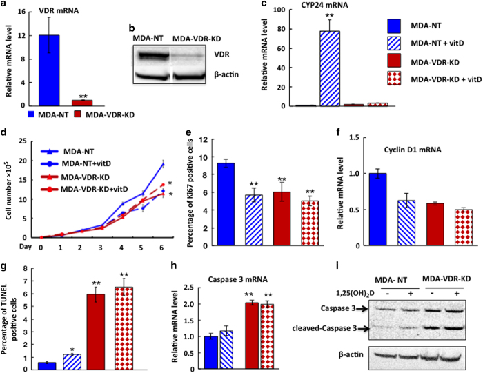
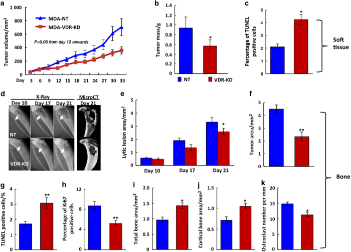
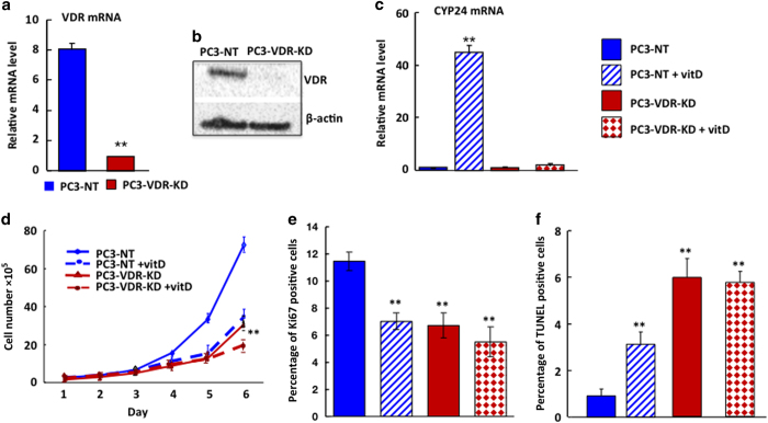
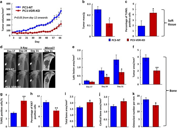
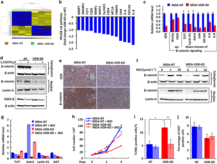
Vitamin D co-regulates cell proliferation, differentiation and apoptosis in numerous tissues, including cancers. The known anti-proliferative and pro-apoptotic actions of the active metabolite of vitamin D, 1,25-dihydroxy-vitamin D [1,25(OH)2D] are mediated through binding to the vitamin D receptor (VDR). Here, we report on the unexpected finding that stable knockdown of VDR expression in the human breast and prostate cancer cell lines, MDA-MB-231 and PC3, strongly induces cell apoptosis and inhibits cell proliferation in vitro. Implantation of these VDR knockdown cells into the mammary fat pad (MDA-MB-231), subcutaneously (PC3) or intra-tibially (both cell lines) in immune-incompetent nude mice resulted in reduced tumor growth associated with increased apoptosis and reduced cell proliferation compared with controls. These growth-retarding effects of VDR knockdown occur in the presence and absence of vitamin D and are independent of whether cells were grown in bone or soft tissues. Transcriptome analysis of VDR knockdown and non-target control cell lines demonstrated that loss of the VDR was associated with significant attenuation in the Wnt/β-catenin signaling pathway. In particular, cytoplasmic and nuclear β-catenin protein levels were reduced with a corresponding downregulation of downstream genes such as Axin2, Cyclin D1, interleukin-6 (IL-6), and IL-8. Stabilization of β-catenin using the GSK-3β inhibitor BIO partly reversed the growth-retarding effects of VDR knockdown. Our results indicate that the unliganded VDR possesses hitherto unknown functions to promote breast and prostate cancer growth, which appear to be operational not only within but also outside the bone environment. These novel functions contrast with the known anti-proliferative nuclear actions of the liganded VDR and may represent targets for new diagnostic and therapeutic approaches in breast and prostate cancer.


Conflict of interest statement

The authors declare no conflict of interest.

Figures

Figure 1
Figure 1
VDR knockdown in MDA-MB-231 cells reduces cell growth in vitro in a ligand-independent manner. (a,b) Compared to non-target controls (MDA-NT, cells transfected with non-target RNA), VDR mRNA (a) and protein (b) expression were knocked down by approximately 80% in MDA-VDR-KD cells at 24 h post plating. **P<0.01. (c) Treatment of MDA-NT and MDA-VDR-KD cells with 10−8 mol·L−1 1,25(OH)2D3 for 8 h increased CYP24 expression by 80-fold in MDA-NT cells with no appreciable response in MDA-VDR-KD cells. **P<0.01 compared to baseline. (d–i) Culture of MDA-NT and MDA-VDR-KD cells under ligand-free conditions: Compared to NT cells, cell growth (d) and cell proliferation (Ki67 immunoreactivity, (e) as well as Cyclin D1 mRNA of VDR-KD cells (f) were reduced by 40%, 36% and 50%, respectively. However, apoptosis was increased 6-fold (g) and Caspase 3 mRNA and protein was increased by 50% (h,i). Treatment of MDA-NT cells with 10−8 mol·L−1 1,25(OH)2D3 reduced both cell growth (d), Ki67 positivity (e) and Cyclin D1 mRNA expression (f) and induced a 2-fold increase in cell apoptosis (g) as well as Caspase 3 mRNA and protein expression (h,i) compared to untreated MDA-NT cells. In contrast, the same treatment had no effect on the growth or apoptosis rates of MDA-VDR-KD cells (d–i). Asterisks denote significant difference from untreated MDA-NT cells (*P<0.05; **P<0.01). In vitro experiments were performed in triplicate and repeated at least three times. Results shown are from a single representative experiment. Data are expressed as mean±s.e.m. (n=3).
Figure 2
Figure 2
VDR knockdown in MDA-MB-231 cells reduces tumor growth in vivo. (a–c) Orthotopic implantation: When implanted into the mammary fat pad, tumors derived from MDA-VDR-KD cells grew significantly slower than those induced by MDA-NT cells (n=10) (a). At study endpoint (day 33 post implantation), tumor weight (b) was reduced by 40% while the proportion of apoptotic cancer cells (c) was increased by 36% in MDA-VDR-KD compared to MDA-NT tumors (n=10). Asterisks denote significant difference from controls (*P<0.05). Data are mean±s.e.m. (d–j) Intra-tibial implantation: At endpoint (day 21 post implantation) lytic lesion size on X-ray, micro-CT (d,e) and histological tumor area (f) were significantly smaller in mice implanted with MDA-VDR-KD than with MDA-NT cells (n=15). Compared to NT controls, tumors derived from VDR knockdown cells were characterized by an increased proportion of apoptotic cancer cells (g) and lower mitotic activity (h). At the bone/tumor interface, tumors derived from VDR knockdown cells exhibited increased total and cortical bone area (i,j), and reduced osteoclast number (k). Asterisks denote significant difference from MDA-NT (*P<0.05; **P<0.01). Data are mean±s.e.m.
Figure 3
Figure 3
Effects of VDR knockdown in prostate cancer (PC3) cells—in vitro studies. (a,b) Compared to non-target controls (PC3-NT, cells transfected with non-target RNA), VDR mRNA (A) and protein (B) expression was knocked down by 80% in PC3-VDR-KD cells at 24 h post plating. **P< 0.001. (c) Treatment of PC3-NT and PC3-VDR-KD cells with 10−8 mol·L−1 1,25(OH)2D3 for 8 h increased CYP24 expression by more than 40-fold in NT cells with no appreciable response in knockdown cells. **P<0.001 compared to baseline. (d-f): Culture of PC3-NT and PC3-VDR-KD cells under ligand-free conditions. Compared to NT cells, cell growth (d) and cell proliferation (Ki67 immunoreactivity, (e) of MDA-VDR-KD cells were reduced by 49% and 41%, respectively, while apoptosis was increased to six fold (f). Treatment of PC3-NT cells with 108 mol·L−1 1,25(OH)2D3 reduced cell growth by 51% (d) and Ki67 positivity by 38% (e) while inducing a 3-fold increase in apoptosis (f) compared to untreated PC3-NT cells. In contrast, the same treatment had no effect on the growth or apoptosis rates of PC3-VDR-KD cells (d–f). *P<0.05; **P<0.01 compared to respective controls. In vitro experiments were performed in triplicate and repeated at least three times. Results shown are from a single representative experiment. Data are expressed as mean±s.e.m. (n=3).
Figure 4
Figure 4
VDR knockdown in PC3 cells reduces tumor growth in vivo. (a–c) Orthotopic implantation (soft tissue, male nude mice). Tumors derived from PC3-VDR-KD cells grew significantly slower than those induced by PC3-NT cells (n=9) (a). At study endpoint (day 69 p.i.) tumor weight (b) was reduced by 40% and the proportion of apoptotic cells (c) was increased by 125% in PC3-VDR-KD compared to PC3-NT tumors (n=10). Asterisks denote significant difference from controls (*P<0.05). Data are mean±s.e.m. (d–j) Intra-tibial implantation: Lytic lesion size (d,e) and tumor area (f) at endpoint (day 31 p. i.) were significantly smaller in mice implanted with PC3-VDR-KD cells compared to those implanted with PC3-NT cells (n=12). Compared to NT controls, tumors derived from PC3-VDR-KD cells were characterized by increased apoptosis (g) and lower mitotic activity (h), and at the bone/tumor interface exhibited increased total and cortical bone area (i,j) and reduced osteoclast number (k). Asterisks denote significance difference from controls (*P<0.05; **P<0.01). Data are mean±s.e.m.
Figure 5
Figure 5
Knockdown of the VDR in MDA-MD-231 breast cancer cells downregulates Wnt/β-catenin signaling pathway. (a) Hierarchical clustering of 121 differentially expressed genes (P<0.05) between MDA-VDR-KD compared to MDA-NT cell lines (microarray data set) showed distinctive expression patterns. The three lanes represent gene array patterns derived from three separate cultures of the same cell line (MD-NT or MDA-VDR-KD) cultured in identical conditions for the same amount of time. (b) From this list, candidate genes belonging to the Wnt/β-catenin pathway, downstream of β-catenin were shown to be downregulated. (c) qRT-PCR was used to validate these genes whereby reflecting processes in decreased proliferation and increased apoptosis. (d,e) Both cytoplasmic and nuclear β-catenin protein levels were reduced in MDA-VDR-KD compared to MDA-NT cells. Treatment with 10−8 mol·L−1 1,25(OH)2D3 induced nuclear β-catenin protein levels in MDA-NT but not in MDA-VDR-KD cells while GSK-3β expression remained unchanged in 1,25(OH)2D3-treated MDA-NT and MDA-VDR-KD cells (d). Reduced β-catenin protein levels in tumors derived from VDR knockdown cells relative to NT controls (IHC stain, e). (f–j) Inhibition of GSK-3β via treatment of VDR-NT and VDR-KD cells with 1 μmol·L−1 BIO resulted in increased expression levels of β-catenin protein (f) as well as Axin2, cyclin D1 and Ki67 mRNA (g). Inhibition of GSK-3β was associated with increased growth of MDA-VDR-KD cells (h), significantly reduced apoptosis (i) but unchanged proliferation of VDR-KD cells, as assessed by Ki67 expression (j). Asterisks denote significance difference from controls (*P<0.05; **P<0.01). Data are mean±s.e.m.

Similar articles

Cited by

References

    1. Robinson BD, Sica GL, Liu YF et al. Tumor microenvironment of metastasis in human breast carcinoma: a potential prognostic marker linked to hematogenous dissemination. Clin Cancer Res 2009; 15: 2433–2441. - PMC - PubMed
    1. Mundy GR. Metastasis to bone: causes, consequences and therapeutic opportunities. Nat Rev Cancer 2002; 2: 584–593. - PubMed
    1. Weilbaecher KN, Guise TA, McCauley LK. Cancer to bone: a fatal attraction. Nat Rev Cancer 2011; 11: 411–425. - PMC - PubMed
    1. Ooi LL, Zheng Y, Stalgis-Bilinski K et al. The bone remodeling environment is a factor in breast cancer bone metastasis. Bone 2011; 48: 66–70. - PubMed
    1. Zheng Y, Zhou H, Dunstan C et al. The role of the bone microenvironment in skeletal metastasis. J Bone Oncol 2013; 2: 47–57. - PMC - PubMed