

Intraocular pressure control of a novel glaucoma drainage device - in vitro and in vivo studies
- PMID: 28944192
- PMCID: PMC5596218
- DOI: 10.18240/ijo.2017.09.04
Intraocular pressure control of a novel glaucoma drainage device - in vitro and in vivo studies
Abstract
Aim: To evaluate the intraocular pressure (IOP) control of an artificial trabeculum drainage system (ATDS), a newly designed glaucoma drainage device, and postoperative complications in normal rabbit eyes.
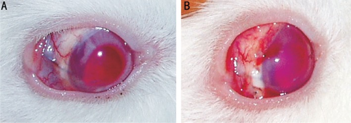
Methods: Pressure drops in air and fluid of 30 ATDS were measured after being connected to a closed manometric system. Twenty of them were then chosen and implanted randomly into the eyes of 20 rabbits. Postoperative slit-lamp, gonioscopic examination and IOP measurements were recorded periodically. Ultrasound biomicroscopy and B-scan ultrasonography were also used to observe the complications. Eyes were enucleated on day 60.
Results: Pressure drops of 4.6-9.4 mm Hg were obtained at physiological aqueous flow rates in the tests in vitro. The average postoperative IOP of the experimental eyes (11.6-12.8 mm Hg) was lower than the controls significantly (P<0.05) at each time point. Complications of hemorrhage (n=1), cellulosic exudation (two cases) and local iris congestion (two cases) were observed. The lumina of the devices were devoid of obstructions in all specimens examined and a thin fibrous capsule was found around the endplate.
Conclusion: ATDS reduce IOP effectively. However, further studies on the structure are needed to reduce complications.
Keywords: aqueous humor; drainage device; intraocular pressure; outflow; rabbit.
Figures


Similar articles
-
A Prospective Analysis of iStent Inject Microstent Positioning: Schlemm Canal Dilatation and Intraocular Pressure Correlations.J Glaucoma. 2019 Jul;28(7):613-621. doi: 10.1097/IJG.0000000000001273. J Glaucoma. 2019. PMID: 31058666 Clinical Trial.
-
[Artificial trabeculum (MESH). Clinical and histological study in the rabbit].J Fr Ophtalmol. 1998 May;21(5):351-60. J Fr Ophtalmol. 1998. PMID: 9759429 French.
-
Aductive laser iridoplasty and laser goniopuncture after non-perforating trabeculectomy.Cesk Slov Oftalmol. 2013 Mar;69(1):3-7. Cesk Slov Oftalmol. 2013. PMID: 23822595
-
Assessment of Filtration Bleb and Endplate Positioning Using Magnetic Resonance Imaging in Eyes Implanted with Long-Tube Glaucoma Drainage Devices.PLoS One. 2015 Dec 7;10(12):e0144595. doi: 10.1371/journal.pone.0144595. eCollection 2015. PLoS One. 2015. PMID: 26641251 Free PMC article.
-
[A challenge to primary open-angle glaucoma including normal-pressure. Clinical problems and their scientific solution].Nippon Ganka Gakkai Zasshi. 2012 Mar;116(3):233-67; discussion 268. Nippon Ganka Gakkai Zasshi. 2012. PMID: 22568103 Review. Japanese.
Cited by
-
International Harmonization of Nomenclature and Diagnostic Criteria (INHAND): Nonproliferative and Proliferative Lesions of Nonrodent Ocular Tissues.Toxicol Pathol. 2024 Oct;52(7):368-455. doi: 10.1177/01926233241283708. Toxicol Pathol. 2024. PMID: 39658869 Free PMC article. Review.
-
Five-in-one: a novel, cost-effective yet simple use of micro needle holder.Int J Ophthalmol. 2022 Apr 18;15(4):657-660. doi: 10.18240/ijo.2022.04.20. eCollection 2022. Int J Ophthalmol. 2022. PMID: 35450178 Free PMC article.
References
-
- Bourne RR, Taylor HR, Flaxman SR, Keeffe J, Leasher J, Naidoo K, Pesudovs K, White RA, Wong TY, Resnikoff S, Jonas JB, Vision Loss Expert Group of the Global Burden of Disease Study Number of people blind or visually impaired by glaucoma worldwide and in world regions 1990 - 2010: a meta-analysis. PLoS One. 2016;11(10):e0162229. - PMC - PubMed
-
- Yu DY, Morgan WH, Sun X, Su EN, Cringle SJ, Yu PK, House P, Guo W, Yu X. The critical role of the conjunctiva in glaucoma filtration surgery. Prog Retin Eye Res. 2009;28(5):303–328. - PubMed
-
- Chaudhry M, Grover S, Baisakhiya S, Bajaj A, Bhatia MS. Artificial drainage devices for glaucoma surgery: an overview. Nepal J Ophthalmol. 2012;4(2):295–302. - PubMed
-
- Wischke C, Neffe AT, Hanh BD, Kreiner CF, Sternberg K, Stachs O, Guthoff RF, Lendlein A. A multifunctional bilayered microstent as glaucoma drainage devices. J Control Release. 2013;172(3):1002–1010. - PubMed
LinkOut - more resources
Full Text Sources
Other Literature Sources