Fetal Heart Sounds Detection Using Wavelet Transform and Fractal Dimension
- PMID: 28944222
- PMCID: PMC5596097
- DOI: 10.3389/fbioe.2017.00049
Fetal Heart Sounds Detection Using Wavelet Transform and Fractal Dimension
Abstract
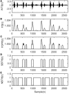
Phonocardiography is a non-invasive technique for the detection of fetal heart sounds (fHSs). In this study, analysis of fetal phonocardiograph (fPCG) signals, in order to achieve fetal heartbeat segmentation, is proposed. The proposed approach (namely WT-FD) is a wavelet transform (WT)-based method that combines fractal dimension (FD) analysis in the WT domain for the extraction of fHSs from the underlying noise. Its adoption in this field stems from its successful use in the fields of lung and bowel sounds de-noising analysis. The efficiency of the WT-FD method in fHS extraction has been evaluated with 19 simulated fHS signals, created for the present study, with additive noise up to (3 dB), along with the simulated fPCGs database available at PhysioBank. Results have shown promising performance in the identification of the correct location and morphology of the fHSs, reaching an overall accuracy of 89% justifying the efficacy of the method. The WT-FD approach effectively extracts the fHS signals from the noisy background, paving the way for testing it in real fHSs and clearly contributing to better evaluation of the fetal heart functionality.
Keywords: fetal heart rate; fetal heart sound; fetal phonocardiogram; fractal dimension thresholding; wavelet transform.
Conflict of interest statement
The authors declare that the research was conducted in the absence of any commercial or financial relationships that could be construed as a potential conflict of interest. The reviewer, WT, and handling editor declared their shared affiliation, and the handling editor states that the process nevertheless met the standards of a fair and objective review.
Figures


Similar articles
-
Detecting fetal heart sounds by means of Fractal Dimension analysis in the Wavelet domain.Annu Int Conf IEEE Eng Med Biol Soc. 2017 Jul;2017:2201-2204. doi: 10.1109/EMBC.2017.8037291. Annu Int Conf IEEE Eng Med Biol Soc. 2017. PMID: 29060333
-
Wavelet filtering of fetal phonocardiography: A comparative analysis.Math Biosci Eng. 2019 Jun 29;16(5):6034-6046. doi: 10.3934/mbe.2019302. Math Biosci Eng. 2019. PMID: 31499751
-
Wavelet-based enhancement of lung and bowel sounds using fractal dimension thresholding--Part II: application results.IEEE Trans Biomed Eng. 2005 Jun;52(6):1050-64. doi: 10.1109/tbme.2005.846717. IEEE Trans Biomed Eng. 2005. PMID: 15977735 Clinical Trial.
-
Analysis of phonocardiogram signals using wavelet transform.J Med Eng Technol. 2012 Aug;36(6):283-302. doi: 10.3109/03091902.2012.684830. Epub 2012 Jun 28. J Med Eng Technol. 2012. PMID: 22738192 Review.
-
Acquisition Devices for Fetal Phonocardiography: A Scoping Review.Bioengineering (Basel). 2024 Apr 11;11(4):367. doi: 10.3390/bioengineering11040367. Bioengineering (Basel). 2024. PMID: 38671788 Free PMC article.
Cited by
-
Hardware/Software Co-design of Fractal Features based Fall Detection System.Sensors (Basel). 2020 Apr 18;20(8):2322. doi: 10.3390/s20082322. Sensors (Basel). 2020. PMID: 32325712 Free PMC article.
-
FPGA-Based High-Performance Phonocardiography System for Extraction of Cardiac Sound Components Using Inverse Delayed Neuron Model.Front Med Technol. 2021 Aug 12;3:666650. doi: 10.3389/fmedt.2021.666650. eCollection 2021. Front Med Technol. 2021. PMID: 35047923 Free PMC article.
-
A comparative study of single-channel signal processing methods in fetal phonocardiography.PLoS One. 2022 Aug 19;17(8):e0269884. doi: 10.1371/journal.pone.0269884. eCollection 2022. PLoS One. 2022. PMID: 35984866 Free PMC article.
-
Beat-to-beat fetal heart rate analysis using portable medical device and wavelet transformation technique.Heliyon. 2022 Dec 22;8(12):e12655. doi: 10.1016/j.heliyon.2022.e12655. eCollection 2022 Dec. Heliyon. 2022. PMID: 36636218 Free PMC article.
-
A Comparative Study on Fetal Heart Rates Estimated from Fetal Phonography and Cardiotocography.Front Physiol. 2017 Oct 17;8:764. doi: 10.3389/fphys.2017.00764. eCollection 2017. Front Physiol. 2017. PMID: 29089896 Free PMC article.
References
-
- Adithya P. C., Sankar R., Moreno W. A., Hart S. (2017). Trends in fetal monitoring through phonocardiography: challenges and future directions. Biomed. Signal Process. Control 33, 289–305.10.1016/j.bspc.2016.11.007 - DOI
LinkOut - more resources
Full Text Sources
Other Literature Sources
Miscellaneous

