. 2017 Jun;4(2):43-63.
doi: 10.1016/j.gendis.2017.04.001. Epub 2017 Apr 27.

Adenovirus-Mediated Gene Delivery: Potential Applications for Gene and Cell-Based Therapies in the New Era of Personalized Medicine

Affiliations

Adenovirus-Mediated Gene Delivery: Potential Applications for Gene and Cell-Based Therapies in the New Era of Personalized Medicine

Cody S Lee et al. Genes Dis. 2017 Jun.

Abstract

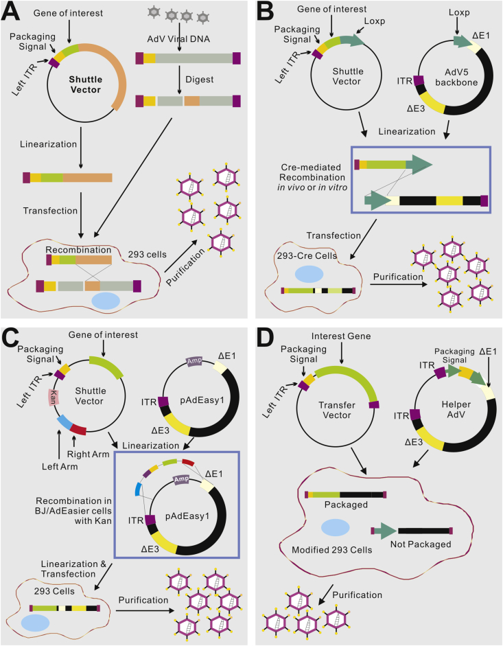
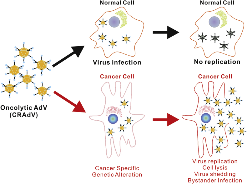
With rapid advances in understanding molecular pathogenesis of human diseases in the era of genome sciences and systems biology, it is anticipated that increasing numbers of therapeutic genes or targets will become available for targeted therapies. Despite numerous setbacks, efficacious gene and/or cell-based therapies still hold the great promise to revolutionize the clinical management of human diseases. It is wildly recognized that poor gene delivery is the limiting factor for most in vivo gene therapies. There has been a long-lasting interest in using viral vectors, especially adenoviral vectors, to deliver therapeutic genes for the past two decades. Among all currently available viral vectors, adenovirus is the most efficient gene delivery system in a broad range of cell and tissue types. The applications of adenoviral vectors in gene delivery have greatly increased in number and efficiency since their initial development. In fact, among over 2,000 gene therapy clinical trials approved worldwide since 1989, a significant portion of the trials have utilized adenoviral vectors. This review aims to provide a comprehensive overview on the characteristics of adenoviral vectors, including adenoviral biology, approaches to engineering adenoviral vectors, and their applications in clinical and pre-clinical studies with an emphasis in the areas of cancer treatment, vaccination and regenerative medicine. Current challenges and future directions regarding the use of adenoviral vectors are also discussed. It is expected that the continued improvements in adenoviral vectors should provide great opportunities for cell and gene therapies to live up to its enormous potential in personalized medicine.

Keywords: adenoviral vector; adenovirus; cell therapy; gene delivery; gene therapy; gene transfer; non-viral vectors; oncolytic virus; personalized medicine; regenerative medicine; tissue engineering; vaccine development; viral vectors.


Conflict of interest statement

CONFLICT OF INTEREST The authors declare no conflicts of interest.

Figures

Figure 1
Figure 1
The genome structure and major transcript units of human Ad5. The Ad5 genome is composed of 36 kb linear double-stranded DNA, also shown in map units (mu). The Ad5 genes are temporally transcribed as early units (E1, E2, E3 and E4 units) or late unit (L1 to L5) in both directions. The top part of the listed genes are transcribed from left to right, while the lower part of the genes are transcribed from right to left, as indicated by the dotted arrows. E1 gene products are involved in the replication of the virus. The E2 region is sub-divided into E2A and E2B. These proteins provide the machinery for viral DNA replication and the ensuing transcription of late genes, which mostly encode structural proteins for virus packaging. Most of the E3 proteins are involved in modulating the immune response of infected cells and are not essential for viral production in vitro.
Figure 2
Figure 2
The structure of adenovirus. (A) Adenovirus is a large, non-enveloped virus presenting icosahedral symmetry. The hexon, penton base, and knobbed fiber, are the most important capsid proteins for gene delivery. (B) Hexon is the major protein forming the 20 triangular faces of the viral capsid. The 240 hexon capsomers in the capsid are trimers, each interacting with six other trimers. The 12 vertices are formed by the penton capsomere, a complex of five copies of the penton base, and three copies of fiber. Each penton capsomere interacts with five hexon capsomeres, one from each of the five faces that converge at the vertex. The knobbed fiber protrudes from the fiber base. In addition, the 5′ termini of adenovirus genome bind covalently to the precursor terminal protein (pTP). The viral genome DNA is wrapped in a histone-like protein and contains the inverted terminal repeats (ITRs), which act as origins of replication.
Figure 3
Figure 3
Adenovirus-mediated gene delivery to mammalian cells. The initial step of adenovirus infection is gaining access to the host cell, and the 12 spikes of the capsid are adhesion receptors, which recognize and bind to specific glycoprotein receptors, such as CAR on the target cell membrane, with the irreversible binding of cell-surface integrins to the penton at the base of the spike. This leads to endocytosis, and the cell-surface membrane invaginates, forming a pit. This pit invaginates and pinches off as a vesicle in the cytoplasm, coated by clathrin and containing the virus. The vesicles are sent to endosome. When the endosome becomes more acidic, the virus is uncoated as the outer capsid disassembles, revealing the viral DNA-protein core. The shed spikes have a toxic function and breach the endosome membrane, allowing the viral core to escape from the endosome into the cytosol of the host cell. Subsequently, the transgenes or nucleic acids are expressed in the target cells.
Figure 4
Figure 4
Three generations of adenoviral vectors. The first generation vectors are deficient in the E1 and E3 regions and have a maximum capacity of 8.2 kb for introducing of a therapeutic gene. The second generation vectors are deficient in E2 and E4, in addition to the deletion of the E1 and E3 regions. Gutless Ad vectors, also known as helper-dependent adenoviruses (HD-Ad), or high-capacity adenoviruses (HC-Ad), can be created to prevent the problem of Ad-created cellular immune response. HD-AdVs are helper-dependent because they depend on a helper adenovirus to be able to produce. HD-AdVs are of high capacity because they allow insertions of up to 36 Kb. Gutless Ad maintain only the 5′ and 3′ ITRs and the packaging signal (Ψ), an essential for final assembly of the virion.
Figure 5
Figure 5
Four commonly-used methods to generate and produce adenovirus vectors for gene delivery. (A) The traditional method – recombination in HEK-293 cells. The gene of interest (GOI) is first cloned into a shuttle vector, which contains 5′-ITR, packaging signal and homologous regions to adenoviral genome. Adenoviruses are generated in HEK-293 cells through recombination between shuttle vector and adenoviral backbone vector, which is unable to produce virus by its self. (B) Cre/LoxP-mediated recombination. The GOI is cloned into a shuttle vector that contains LoxP site(s). Cre recombinase-mediated recombination occurs with a LoxP-containing adenoviral backbone vector in vitro or 293-Cre cells, leading to the generation of adenoviruses. (C) The AdEasy system. The GOI is subcloned into a shuttle vector that contains 5′-ITR and packaging signal, as well as a kanamycin-containing bacterial replication unit flanked with homologous arms. Recombinant adenoviral plasmids are generated through homologous recombination between the linearized shuttle vector and ampicillin-resistant adenoviral backbone vector, such as pAdEasy1, in the bacterial strain BJ5183 cells under kanamycin selection. The resultant adenoviral plasmids are linearized and used for adenovirus production in HEK-293 cells. (D) The use of helper adenovirus for the production of HC-AdVs (or HD-AdVs, or Gutless AdVs). The GOI is cloned into a transfer vector that contains both ITRs and packaging signal only. Adenoviruses are generated with a helper adenovirus, which will not be packaged due to the deletion of packaging signal in the modified HEK-293 cells, usually through Cre/LoxP or FLP/FRT excision system.
Figure 6
Figure 6
The anticancer effect of oncolytic adenoviruses (CRAdVs). The oncolytic adenoviruses infect both normal and cancer cells with similar efficiency. In cancer cells cancer-specific genetic changes activate the expression of Ad5 E1 genes, which initiates viral replication cascade, leading to virus production and cancer cell lysis. The packaged viruses are released and infect neighboring cancer cells to achieve bystander killing effect. However, normal cells lack the cancer-specific activation of viral replication, and thus no Ad5 viruses are reproduced. As the result, normal cells are spared by oncolytic adenoviruses.

Similar articles

Cited by

References

    1. Kay M.A., Glorioso J.C., Naldini L. Viral vectors for gene therapy: the art of turning infectious agents into vehicles of therapeutics. Nat Med. 2001;7(1):33–40. - PubMed
    1. Crystal R.G. Adenovirus: the first effective in vivo gene delivery vector. Hum Gene Ther. 2014;25(1):3–11. - PMC - PubMed
    1. Breyer B., Jiang W., Cheng H. Adenoviral vector-mediated gene transfer for human gene therapy. Curr Gene Ther. 2001;1(2):149–162. - PubMed
    1. Seymour L.W., Fisher K.D. Adenovirus: teaching an old dog new tricks. Hum Gene Ther. 2011;22(9):1041–1042. - PubMed
    1. Rauschhuber C., Noske N., Ehrhardt A. New insights into stability of recombinant adenovirus vector genomes in mammalian cells. Eur J Cell Biol. 2012;91(1):2–9. - PubMed