. 2017 Aug 4;1(1):2398212817723443.
doi: 10.1177/2398212817723443.

Hippocampal - diencephalic - cingulate networks for memory and emotion: An anatomical guide

Affiliations

Hippocampal - diencephalic - cingulate networks for memory and emotion: An anatomical guide

Emma J Bubb et al. Brain Neurosci Adv. .

Abstract

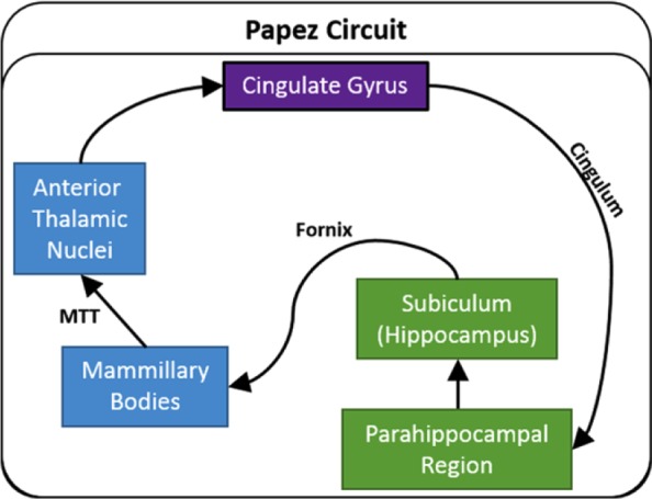
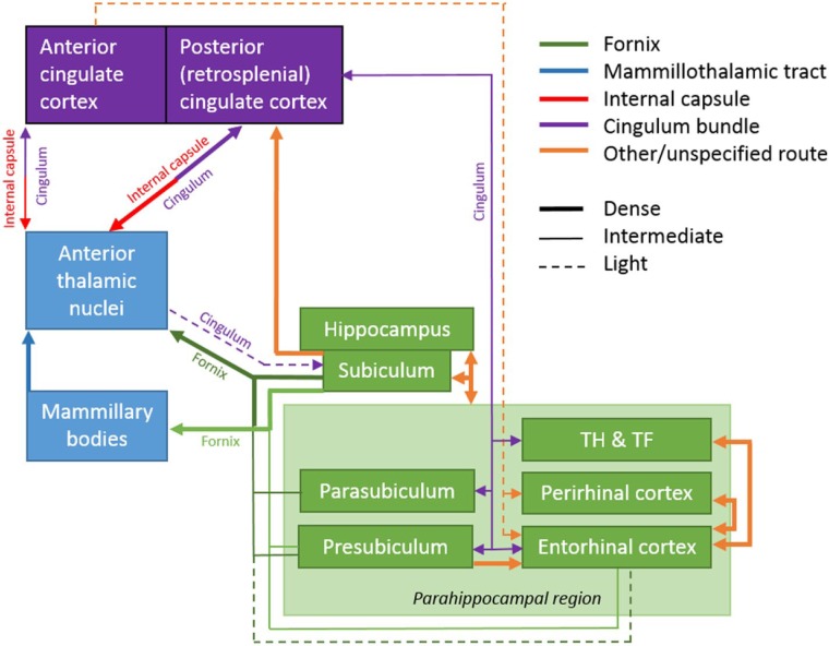
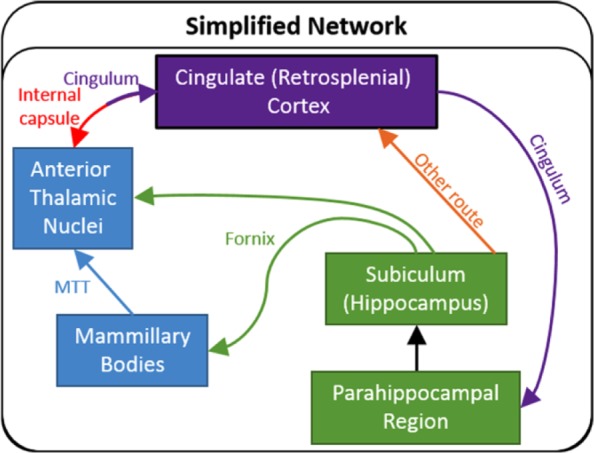
This review brings together current knowledge from tract tracing studies to update and reconsider those limbic connections initially highlighted by Papez (1937) for their presumed role in emotion. These connections link hippocampal and parahippocampal regions with the mammillary bodies, the anterior thalamic nuclei, and the cingulate gyrus, all structures now strongly implicated in memory functions. An additional goal of this review is to describe the routes taken by the various connections within this network. The original descriptions of these limbic connections saw their interconnecting pathways forming a serial circuit that began and finished in the hippocampal formation. It is now clear that, with the exception of the mammillary bodies, these various sites are multiply interconnected with each other, including many reciprocal connections. In addition, these same connections are topographically organised, creating further subsystems. This complex pattern of connectivity helps to explain the difficulty of interpreting the functional outcome of damage to any individual site within the network. For these same reasons, Papez' initial concept of a loop beginning and ending in the hippocampal formation needs to be seen as a much more complex system of hippocampal-diencephalic-cingulate connections. The functions of these multiple interactions might be better viewed as principally providing efferent information from the posterior medial temporal lobe. Both a subcortical diencephalic route (via the fornix) and a cortical cingulate route (via retrosplenial cortex) can be distinguished. These routes provide indirect pathways for hippocampal interactions with prefrontal cortex, with the preponderance of both sets of connections arising from the more posterior hippocampal regions. These multi-stage connections complement the direct hippocampal projections to prefrontal cortex, which principally arise from the anterior hippocampus, thereby creating longitudinal functional differences along the anterior-posterior plane of the hippocampus.

Keywords: Cingulate cortex; Cingulum; Fornix; Hippocampus; Mammillary bodies; Papez circuit; Parahippocampal cortex; Review; Subiculum; Thalamus.


Conflict of interest statement

Declaration of Conflicting Interests: The Authors declare that there is no conflict of interest.

Figures

Figure 1.
Figure 1.
Traditional depiction of Papez circuit. The arrows show the direction of each set of connections. MTT: mammillothalamic tract.
Figure 2.
Figure 2.
Nissl-stained coronal sections from the rat (left column) and macaque monkey (right column) showing most of the structures that comprise the hippocampal–diencephalic–cingulate network. The anterior thalamic nuclei are at the top, the hippocampus and parahippocampal region are in the middle, while the mammillary bodies are at the bottom. Left: The labels ‘Deep–Superficial’, ‘Distal–Proximal’, and ‘Septal–Temporal’ depict the three planes within the hippocampus. Scale bars = 500 µm. Right: Sections from a monkey (Macaca fascicularis). Scale bars = 1000 µm. AD: anterodorsal nucleus; AM: anteromedial nucleus; AV: anteroventral nucleus; CA1–3: CA fields of the hippocampus; DG: dentate gyrus; LM: lateral nucleus of the mammillary bodies; MM: medial nucleus of the mammillary bodies; MML: lateral division of the medial mammillary nucleus; MMM: medial division of the medial mammillary nucleus; Para: parasubiculum; Post: postsubiculum; Pre: presubiculum; Pro: prosubiculum; Rdg: dysgranular retrosplenial cortex (area 30); Rga: Rgb: subregions within granular retrosplenial cortex (area 29); Sub: subiculum. Note: parahippocampal areas TH and TF (and postrhinal cortex) are not depicted, neither are the monkey cingulate cortices as this would involve additional planes (see Figure 8).
Figure 3.
Figure 3.
The rat brain. Depiction of the connections between the hippocampal and parahippocampal regions with the mammillary bodies and anterior thalamic nuclei, as well as the projections from the mammillary bodies to the anterior thalamic nuclei. The routes of these connections are distinguished by different colours. The origin of a connection is denoted by a circle and the termination is signified by an arrowhead, while a reciprocal connection that follows the same route has an arrowhead at both ends. The style of the lines reflects the strength of the connections (thick line = dense, thin line = intermediate, dashed line = light). The upper left panel shows the anterior thalamic nuclei, while the lower left panel depicts the mammillary bodies. AD: anterodorsal nucleus; AM: anteromedial nucleus; AV: anteroventral nucleus; CA1–3: CA fields of the hippocampus; DG: dentate gyrus; LM: lateral nucleus of the mammillary bodies; MM: medial nucleus of the mammillary bodies; MML: lateral division of the medial mammillary nucleus; MMM: medial division of the medial mammillary nucleus; Post: postsubiculum; Pre: presubiculum; PRH: perirhinal cortex; Rdg: dysgranular retrosplenial cortex (area 30); Rga: Rgb: subregions within granular retrosplenial cortex (area 29); Sub: subiculum. Scale bar = 500 µm.
Figure 4.
Figure 4.
The rat brain. Depiction of the connections between the anterior thalamic nuclei (lower left), cingulate cortices, hippocampus, and parahippocampal areas. The routes of these projections are distinguished by different colours. The origin of a connection is denoted by a circle and the termination is signified by an arrowhead while a reciprocal connection that follows the same route has an arrowhead at both ends. The style of the lines reflects the strength of the connections (thick line = dense, thin line = intermediate, dashed line = light). AD: anterodorsal nucleus; AM: anteromedial nucleus; AV: anteroventral nucleus; CA1–3: CA fields of the hippocampus; CC: corpus callosum; DG: dentate gyrus; Post: postsubiculum; Pre: presubiculum; PRH: perirhinal cortex; Rdg: dysgranular retrosplenial cortex (area 30); Rga: Rgb: subregions within granular retrosplenial cortex (area 29); Sub: subiculum. Scale bar = 500 µm.
Figure 5.
Figure 5.
Schematic showing the major hippocampal–parahippocampal interactions in the rat brain. These interconnections are organised by both their proximal–distal locations and the lamina of their inputs and outputs. CA: CA fields of the hippocampus; Dist: distal; DG: dentate gyrus; Prox: proximal; Sub: subiculum. (For simplicity, the presubiculum and parasubiculum are not included.)
Figure 6.
Figure 6.
The rat brain: Schematic showing the main, direct interconnections between sites in Papez circuit. The style of the lines reflects the strength of the connections (thick line = dense, thin line = intermediate, dashed line = light).
Figure 7.
Figure 7.
Macaque monkey brain. Depiction of the connections between the hippocampal and parahippocampal regions with the mammillary bodies and anterior thalamic nuclei, as well as the projections from the mammillary bodies to the anterior thalamic nuclei. The routes of these connections are distinguished by different colours. The origin of a connection is denoted by a circle and the termination is signified by an arrowhead while a reciprocal connection that follows the same route has an arrowhead at both ends. The style of the lines reflects the strength of the connections (thick line = dense, thin line = intermediate, dashed line = light). The upper left panel shows the anterior thalamic nuclei, while the lower left panel depicts the mammillary bodies. AD: anterodorsal nucleus; AM: anteromedial nucleus; AV: anteroventral nucleus; CA1–3: CA fields of the hippocampus; DG: dentate gyrus; LM: lateral nucleus of the mammillary bodies; MM: medial nucleus of the mammillary bodies; MML: lateral division of the medial mammillary nucleus; MMM: medial division of the medial mammillary nucleus; Para: parasubiculum; Pre: presubiculum; Sub: subiculum. Scale bars = 1000 µm.
Figure 8.
Figure 8.
Macaque monkey brain. Depiction of the connections between the anterior thalamic nuclei (lower left), cingulate gyrus (areas 23, 24, 25, 29, 30, 31, 32), hippocampus, and parahippocampal regions. The routes of these projections are distinguished by different colours. In the case of some connections, two colours are used to show how they pass from other pathway to another. The origin of a connection is denoted by a circle and the termination is signified by an arrowhead while a reciprocal connection that follows the same route has an arrowhead at both ends. AD: anterodorsal nucleus; AM: anteromedial nucleus; AV: anteroventral nucleus; CA1–3: CA fields of the hippocampus; DG: dentate gyrus; Para: parasubiculum; Pre: presubiculum; Sub: subiculum. Scale bars = 1000 µm.
Figure 9.
Figure 9.
Schematic showing the major interconnections of the hippocampal–diencephalic–cingulate network in the macaque monkey. In the case of some connections, two colours are used to show how they pass from one pathway to another. The style of the lines reflects the strength of the connections (thick line = dense, thin line = intermediate, dashed line = light).
Figure 10.
Figure 10.
Simplified schematic showing the major common connections in both the rat and monkey (macaque) brains between those sites comprising the hippocampal–diencephalic–cingulate network. When updated, it is apparent that Papez ‘circuit’ can also be interpreted as twin routes (dorsal and ventral) from the hippocampal and parahippocampal regions to the cingulate cortices and thalamus. The twin colour of the pathway between the anterior thalamic nuclei and the cingulate cortex reflects the involvement of two tracts. MTT: mammillothalamic tract.

References

    1. Aggleton JP. (1986) A description of the amygdalo-hippocampal interconnections in the macaque monkey. Experimental Brain Research 64(3): 515–526. - PubMed
    1. Aggleton JP. (2012) Multiple anatomical systems embedded within the primate medial temporal lobe: Implications for hippocampal function. Neuroscience and Biobehavioral Reviews 36(7): 1579–1596. - PubMed
    1. Aggleton JP, Brown MW. (1999) Episodic memory, amnesia, and the hippocampal-anterior thalamic axis. Behavioral and Brain Sciences 22(3): 425–444. - PubMed
    1. Aggleton JP, Brown MW. (2006) Interleaving brain systems for episodic and recognition memory. Trends in Cognitive Sciences 10(10): 455–463. - PubMed
    1. Aggleton JP, Desimone R, Mishkin M. (1986) The origin, course, and termination of the hippocampothalamic projections in the macaque. Journal of Comparative Neurology 243(3): 409–421. - PubMed

LinkOut - more resources