Proteomic analysis of extracellular vesicles from a Plasmodium falciparum Kenyan clinical isolate defines a core parasite secretome
- PMID: 28944300
- PMCID: PMC5583745
- DOI: 10.12688/wellcomeopenres.11910.2
Proteomic analysis of extracellular vesicles from a Plasmodium falciparum Kenyan clinical isolate defines a core parasite secretome
Abstract
Background: Many pathogens secrete effector molecules to subvert host immune responses, to acquire nutrients, and/or to prepare host cells for invasion. One of the ways that effector molecules are secreted is through extracellular vesicles (EVs) such as exosomes. Recently, the malaria parasite P. falciparum has been shown to produce EVs that can mediate transfer of genetic material between parasites and induce sexual commitment. Characterizing the content of these vesicles may improve our understanding of P. falciparum pathogenesis and virulence.
Methods: Previous studies of P. falciparum EVs have been limited to long-term adapted laboratory isolates. In this study, we isolated EVs from a Kenyan P. falciparum clinical isolate adapted to in vitro culture for a short period and characterized their protein content by mass spectrometry (data are available via ProteomeXchange, with identifier PXD006925).
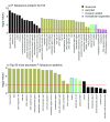
Results: We show that P. falciparum extracellular vesicles ( PfEVs) are enriched in proteins found within the exomembrane compartments of infected erythrocytes such as Maurer's clefts (MCs), as well as the secretory endomembrane compartments in the apical end of the merozoites, suggesting that these proteins play a role in parasite-host interactions. Comparison of this novel clinically relevant dataset with previously published datasets helps to define a core secretome present in Plasmodium EVs.
Conclusions: P. falciparum extracellular vesicles contain virulence-associated parasite proteins. Therefore, analysis of PfEVs contents from a range of clinical isolates, and their functional validation may improve our understanding of the virulence mechanisms of the parasite, and potentially identify targets for interventions or diagnostics.
Keywords: Malaria; Plasmodium falciparum; exosomes; extracellular vesicles; proteomics.
Conflict of interest statement
Competing interests: No competing interest were disclosed
Figures


Similar articles
-
Extracellular Vesicles Derived from Early and Late Stage Plasmodium falciparum-Infected Red Blood Cells Contain Invasion-Associated Proteins.J Clin Med. 2022 Jul 21;11(14):4250. doi: 10.3390/jcm11144250. J Clin Med. 2022. PMID: 35888014 Free PMC article.
-
Extracellular vesicles from early stage Plasmodium falciparum-infected red blood cells contain PfEMP1 and induce transcriptional changes in human monocytes.Cell Microbiol. 2018 May;20(5):e12822. doi: 10.1111/cmi.12822. Epub 2018 Feb 15. Cell Microbiol. 2018. PMID: 29349926
-
Role of Plasmodium falciparum Protein GEXP07 in Maurer's Cleft Morphology, Knob Architecture, and P. falciparum EMP1 Trafficking.mBio. 2020 Mar 17;11(2):e03320-19. doi: 10.1128/mBio.03320-19. mBio. 2020. PMID: 32184257 Free PMC article.
-
Maurer's clefts, the enigma of Plasmodium falciparum.Proc Natl Acad Sci U S A. 2013 Dec 10;110(50):19987-94. doi: 10.1073/pnas.1309247110. Epub 2013 Nov 27. Proc Natl Acad Sci U S A. 2013. PMID: 24284172 Free PMC article. Review.
-
Host cell remodeling by pathogens: the exomembrane system in Plasmodium-infected erythrocytes.FEMS Microbiol Rev. 2016 Sep;40(5):701-21. doi: 10.1093/femsre/fuw016. FEMS Microbiol Rev. 2016. PMID: 27587718 Free PMC article. Review.
Cited by
-
Overview of extracellular vesicles in pathogens with special focus on human extracellular protozoan parasites.Mem Inst Oswaldo Cruz. 2024 Sep 23;119:e240073. doi: 10.1590/0074-02760240073. eCollection 2024. Mem Inst Oswaldo Cruz. 2024. PMID: 39319874 Free PMC article. Review.
-
An update on cerebral malaria for therapeutic intervention.Mol Biol Rep. 2022 Nov;49(11):10579-10591. doi: 10.1007/s11033-022-07625-5. Epub 2022 Jun 7. Mol Biol Rep. 2022. PMID: 35670928 Review.
-
Interaction Analysis of a Plasmodium falciparum PHISTa-like Protein and PfEMP1 Proteins.Front Microbiol. 2020 Nov 13;11:611190. doi: 10.3389/fmicb.2020.611190. eCollection 2020. Front Microbiol. 2020. PMID: 33281807 Free PMC article.
-
The use of proteomics for the identification of promising vaccine and diagnostic biomarkers in Plasmodium falciparum.Parasitology. 2020 Oct;147(12):1255-1262. doi: 10.1017/S003118202000102X. Epub 2020 Jun 19. Parasitology. 2020. PMID: 32618524 Free PMC article. Review.
-
Extracellular Proteomic Profiling from the Erythrocytes Infected with Plasmodium Falciparum 3D7 Holds Promise for the Detection of Biomarkers.Protein J. 2024 Aug;43(4):819-833. doi: 10.1007/s10930-024-10212-1. Epub 2024 Jul 15. Protein J. 2024. PMID: 39009910
References
-
- WHO: World Malaria Report.2016. Reference Source
Grants and funding
LinkOut - more resources
Full Text Sources
Other Literature Sources

