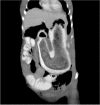
Closed-perforation of gastric fundus and gastric outlet obstruction caused by a giant gastric trichobezoar: A case report
- PMID: 28944343
- PMCID: PMC5602322
- DOI: 10.5152/UCD.2015.2994
Closed-perforation of gastric fundus and gastric outlet obstruction caused by a giant gastric trichobezoar: A case report
Abstract
A bezoar is a mass formed because of the accumulation of indigestible material in the stomach and/or small intestine. Bezoars are rare but occasionally occur with acute abdomen findings. Bezoars form as a result of changes in the gastrointestinal system anatomy and physiology and repetitive exposure to the ingested material. These materials can include vegetables with high fiber content (phytobezoars), non-animal origin fats, hair (trichobezoars), or drugs such as anti-acids (pharmobezoars). Gastric bezoars frequently occur after gastric surgery. Psychiatric disorders such as trichotillomania (an irresistible urge to remove and swallow one's own hair) are frequently the underlying reason in patients without a history of gastric surgery. In this article, we presented a giant gastric trichobezoar obstructing outlet and causing closed-perforation and abscess formation of gastric fundus in a 30-year-old woman.
Keywords: Bezoar; closed-perforation of stomach; gastric outlet obstruction; trichobezoar.
Conflict of interest statement
Conflict of Interest: No conflict of interest was declared by the authors.
Figures


Similar articles
-
A Giant Trichobezoar Causing Rapunzel Syndrome in a 12-year-old Female.Indian J Psychol Med. 2011 Jan;33(1):77-9. doi: 10.4103/0253-7176.85401. Indian J Psychol Med. 2011. PMID: 22021959 Free PMC article.
-
Gastric trichobezoar presenting as gastric outlet obstruction--a case report.Nepal Med Coll J. 2007 Mar;9(1):67-9. Nepal Med Coll J. 2007. PMID: 17593683
-
[Trichobezoar-Rapunzel syndrome--case report].Rozhl Chir. 2004 Sep;83(9):460-2. Rozhl Chir. 2004. PMID: 15615345 Czech.
-
Endoscopic shaving of hair in a gastric bypass patient with a large bezoar.BMJ Case Rep. 2017 Oct 9;2017:bcr2017220923. doi: 10.1136/bcr-2017-220923. BMJ Case Rep. 2017. PMID: 28993354 Free PMC article. Review.
-
Review of the diagnosis and management of gastrointestinal bezoars.World J Gastrointest Endosc. 2015 Apr 16;7(4):336-45. doi: 10.4253/wjge.v7.i4.336. World J Gastrointest Endosc. 2015. PMID: 25901212 Free PMC article. Review.
References
-
- Gurses N, Ozkan K. Bezoars analysis of seven cases. Z Kinderchir. 1987;42:291–292. https://doi.org/10.1055/s-2008-1075605. - DOI - PubMed
-
- Edelstein MM, Freed E, Wexler M. Diospyrobezoar of the jejunum in a post gastrectomy patient. Arch Surg. 1971;103:765–766. https://doi.org/10.1001/archsurg.1971.01350120129026. - DOI - PubMed
-
- Wadlington WB, Rose M, Holcomb GW. Complication of trichobezoars: A 30 year experience. South Med J. 1992;85:1020–1022. https://doi.org/10.1097/00007611-199210000-00024. - DOI - PubMed
-
- Byrme WJ. Foreign bodies, bezoars and caustic ingestion. Gastrointest Endosc Clin North Am. 1994;4:99–144. - PubMed
-
- Tsou JM, Bishop PR, Nowicki MJ. Colonic sunflower seed bezoar. Pediatrics. 1997;99:896–897. https://doi.org/10.1542/peds.99.6.896. - DOI - PubMed
LinkOut - more resources
Full Text Sources
Other Literature Sources
