

Subcellular compartmentalisation of copper, iron, manganese, and zinc in the Parkinson's disease brain
- PMID: 28944802
- PMCID: PMC5647261
- DOI: 10.1039/c7mt00244k
Subcellular compartmentalisation of copper, iron, manganese, and zinc in the Parkinson's disease brain
Abstract
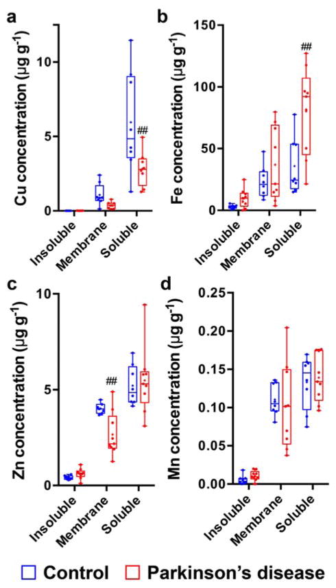
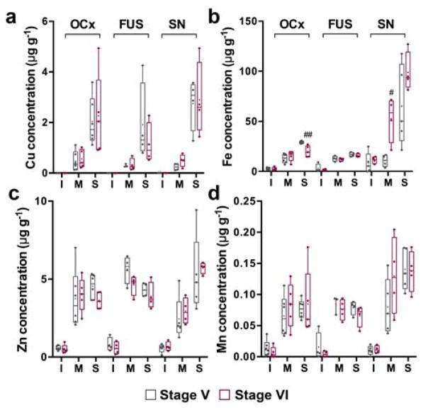
Elevated iron and decreased copper levels are cardinal features of the degenerating substantia nigra pars compacta in the Parkinson's disease brain. Both of these redox-active metals, and fellow transition metals manganese and zinc, are found at high concentrations within the midbrain and participate in a range of unique biological reactions. We examined the total metal content and cellular compartmentalisation of manganese, iron, copper and zinc in the degenerating substantia nigra, disease-affected but non-degenerating fusiform gyrus, and unaffected occipital cortex in the post mortem Parkinson's disease brain compared with age-matched controls. An expected increase in iron and a decrease in copper concentration was isolated to the soluble cellular fraction, encompassing both interstitial and cytosolic metals and metal-binding proteins, rather than the membrane-associated or insoluble fractions. Manganese and zinc levels did not differ between experimental groups. Altered Fe and Cu levels were unrelated to Braak pathological staging in our cases of late-stage (Braak stage V and VI) disease. The data supports our hypothesis that regional alterations in Fe and Cu, and in proteins that utilise these metals, contribute to the regional selectively of neuronal vulnerability in this disorder.
Conflict of interest statement
B.R.R. and D.J.H. receive material support from Agilent Technologies.
Figures

References
-
- Que EL, Domaille DW, Chang CJ. Chem Rev. 2008;108:4328–4328. - PubMed
-
- Chang CJ. Nat Chem Biol. 2015;11:744–747. - PubMed
-
- Dexter DT, Carayon A, Javoy-Agid F, Agid Y, Wells FR, Daniel SE, Lees AJ, Jenner P, Marsden CD. Brain. 1991;114:1953–1975. - PubMed
-
- Barnham KJ, Bush AI. Curr Opin Chem Biol. 2008;12:222–228. - PubMed
Publication types
MeSH terms
Substances
Grants and funding
LinkOut - more resources
Full Text Sources
Other Literature Sources
Medical
Miscellaneous
