

Triptolide inhibits proliferation, differentiation and induces apoptosis of osteoblastic MC3T3‑E1 cells
- PMID: 28944904
- PMCID: PMC5865870
- DOI: 10.3892/mmr.2017.7568
Triptolide inhibits proliferation, differentiation and induces apoptosis of osteoblastic MC3T3‑E1 cells
Abstract
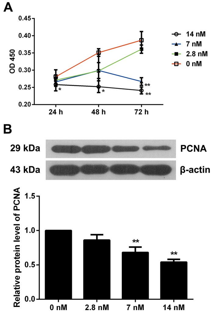
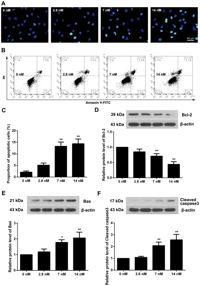
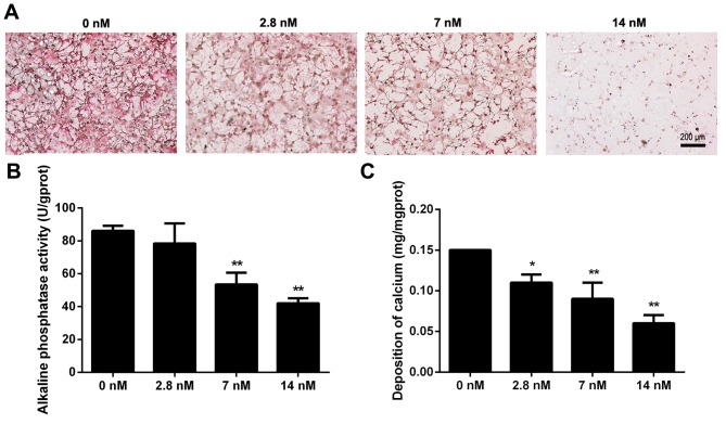
Ankylosing spondylitis (AS) is characterized by the formation of bony spurs. Treatment of the resulting ankylosis, excessive bone formation and associated functional impairment, remain the primary therapeutic aims in research regarding this condition. Triptolide is the primary active component of the perennial vine Tripterygium wilfordii Hook. f., and has previously been demonstrated to exert anti‑tumor activities including inhibition of cell growth and the induction of apoptosis, however, the effect of triptolide on osteoblasts remains to be elucidated. In the present study, the MC3T3‑E1 mouse osteoblast cell line was treated with differing concentrations of triptolide for various intervals. Cell proliferation was detected using the bromodeoxyuridine assay, cell cycle and apoptosis were measured by flow cytometry, nuclear apoptosis was observed by Hoechst staining and associated proteins were determined via western blot analysis. The cells were then further incubated with osteogenic induction medium supplemented with triptolide for 7 or 12 days and the differentiation to osteoblasts was examined by picrosirius staining, observation of alkaline phosphatase activity and a calcium deposition assay. It was demonstrated that treatment with triptolide significantly inhibited osteoblast proliferation and induced cell cycle arrest and apoptosis of the osteoblasts. Furthermore, treatment with triptolide reduced collagen formation, alkaline phosphatase activity and calcium deposition. The present study demonstrated an inhibitory effect of triptolide on osteoblast proliferation and differentiation, and therefore suggests a potential therapeutic agent for the treatment of AS in the future.
Figures

Similar articles
-
Triptolide inhibits cell proliferation and tumorigenicity of human neuroblastoma cells.Mol Med Rep. 2015 Feb;11(2):791-6. doi: 10.3892/mmr.2014.2814. Epub 2014 Oct 29. Mol Med Rep. 2015. PMID: 25354591 Free PMC article.
-
Triptolide induced cell death through apoptosis and autophagy in murine leukemia WEHI-3 cells in vitro and promoting immune responses in WEHI-3 generated leukemia mice in vivo.Environ Toxicol. 2017 Feb;32(2):550-568. doi: 10.1002/tox.22259. Epub 2016 Mar 18. Environ Toxicol. 2017. PMID: 26990902
-
Caspase 3 is involved in the apoptosis induced by triptolide in HK-2 cells.Toxicol In Vitro. 2009 Jun;23(4):598-602. doi: 10.1016/j.tiv.2009.01.021. Epub 2009 Feb 20. Toxicol In Vitro. 2009. PMID: 19233258
-
[Progress in research on mechanisms of anti-rheumatoid arthritis of triptolide].Zhongguo Zhong Yao Za Zhi. 2006 Oct;31(19):1575-9. Zhongguo Zhong Yao Za Zhi. 2006. PMID: 17165577 Review. Chinese.
-
Triptolide and its expanding multiple pharmacological functions.Int Immunopharmacol. 2011 Mar;11(3):377-83. doi: 10.1016/j.intimp.2011.01.012. Epub 2011 Jan 19. Int Immunopharmacol. 2011. PMID: 21255694 Review.
Cited by
-
Human cells with osteogenic potential in bone tissue research.Biomed Eng Online. 2023 Apr 3;22(1):33. doi: 10.1186/s12938-023-01096-w. Biomed Eng Online. 2023. PMID: 37013601 Free PMC article. Review.
-
Magnetic Hydroxyapatite Composite Nanoparticles for Augmented Differentiation of MC3T3-E1 Cells for Bone Tissue Engineering.Mar Drugs. 2023 Jan 25;21(2):85. doi: 10.3390/md21020085. Mar Drugs. 2023. PMID: 36827126 Free PMC article.
-
Triptolide alleviates the development of inflammation in ankylosing spondylitis via the NONHSAT227927.1/JAK2/STAT3 pathway.Exp Ther Med. 2023 Nov 16;27(1):17. doi: 10.3892/etm.2023.12305. eCollection 2024 Jan. Exp Ther Med. 2023. PMID: 38223328 Free PMC article.
-
Therapeutic potential of triptolide in autoimmune diseases and strategies to reduce its toxicity.Chin Med. 2021 Nov 7;16(1):114. doi: 10.1186/s13020-021-00525-z. Chin Med. 2021. PMID: 34743749 Free PMC article. Review.
-
Triptolide attenuates inhibition of ankylosing spondylitis-derived mesenchymal stem cells on the osteoclastogenesis through modulating exosomal transfer of circ-0110634.J Orthop Translat. 2022 Sep 16;36:132-144. doi: 10.1016/j.jot.2022.05.007. eCollection 2022 Sep. J Orthop Translat. 2022. PMID: 36185580 Free PMC article.
References
MeSH terms
Substances
LinkOut - more resources
Full Text Sources
Other Literature Sources
Research Materials
