. 2017 Nov 1;127(11):3923-3936.
doi: 10.1172/JCI95442. Epub 2017 Sep 25.

Mutations in the netrin-1 gene cause congenital mirror movements

Affiliations

Mutations in the netrin-1 gene cause congenital mirror movements

Aurélie Méneret et al. J Clin Invest. .

Abstract

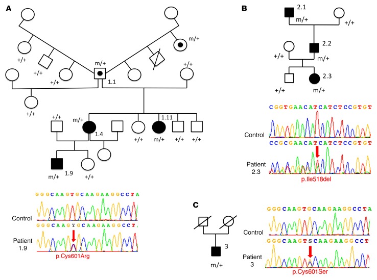
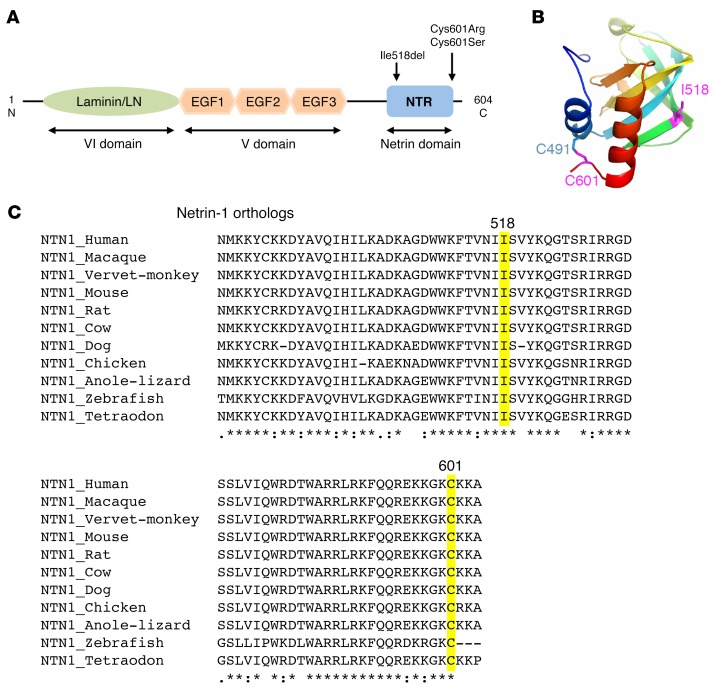
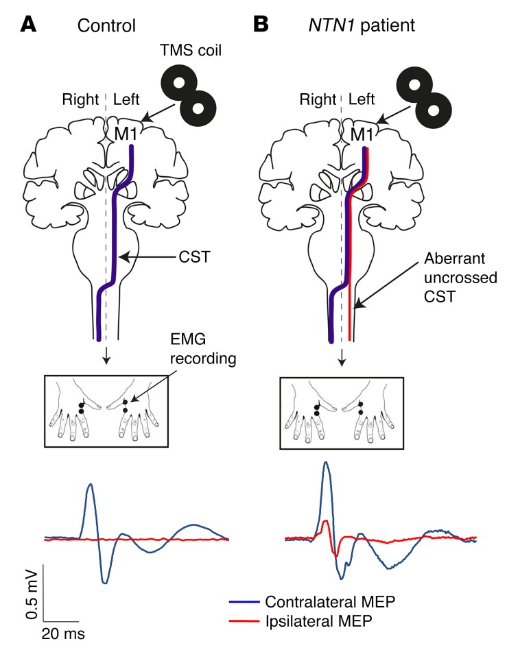
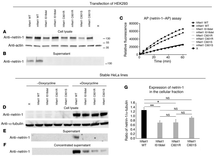
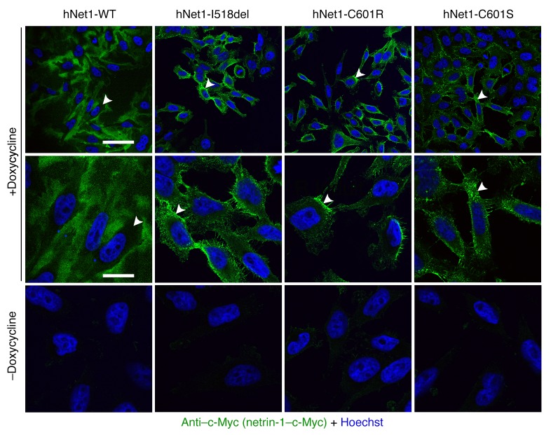
Netrin-1 is a secreted protein that was first identified 20 years ago as an axon guidance molecule that regulates midline crossing in the CNS. It plays critical roles in various tissues throughout development and is implicated in tumorigenesis and inflammation in adulthood. Despite extensive studies, no inherited human disease has been directly associated with mutations in NTN1, the gene coding for netrin-1. Here, we have identified 3 mutations in exon 7 of NTN1 in 2 unrelated families and 1 sporadic case with isolated congenital mirror movements (CMM), a disorder characterized by involuntary movements of one hand that mirror intentional movements of the opposite hand. Given the diverse roles of netrin-1, the absence of manifestations other than CMM in NTN1 mutation carriers was unexpected. Using multimodal approaches, we discovered that the anatomy of the corticospinal tract (CST) is abnormal in patients with NTN1-mutant CMM. When expressed in HEK293 or stable HeLa cells, the 3 mutated netrin-1 proteins were almost exclusively detected in the intracellular compartment, contrary to WT netrin-1, which is detected in both intracellular and extracellular compartments. Since netrin-1 is a diffusible extracellular cue, the pathophysiology likely involves its loss of function and subsequent disruption of axon guidance, resulting in abnormal decussation of the CST.


Conflict of interest statement

Conflict of interest: The authors have declared that no conflict of interest exists.

Figures

Figure 1
Figure 1. Pedigrees of the CMM families and distribution of the mutations in NTN1.
(A) Family 1 (from France) with a C601R mutation (the unusual family tree is a result of individual 1.1, who had children with 4 different women), (B) family 2 (from the United Kingdom [data were collected in New Zealand], which was previously identified as Family C in ref. 33), with an I518del mutation, and (C) a Canadian sporadic case with a C601S mutation. Black symbols represent individuals with CMM, symbols with an embedded black circle indicate asymptomatic carriers, white symbols indicate unaffected individuals, and symbols with a diagonal line represent deceased individuals. Squares represent males and circles represent females. m, mutated allele; +, WT allele. Electrophoregrams confirm the 3 NTN1 mutations obtained by Sanger sequencing. Red arrows point to sequence changes. Electrophoregrams were obtained with SeqScape software, version 2.6 (Applied Biosystems).
Figure 2
Figure 2. Position and conservation of the mutated amino acids.
(A) Schematic of the netrin-1 protein showing the laminin (LN) domain, the 3 EGF-like domains, and the netrin (NTR) domain. Arrows show the location of the 3 mutations. (B) Structural model of the NTR domain of WT netrin-1 showing the 2 cysteines involved in a disulfide bridge (C601 and C491) and I518, which has a key position in a β-strand. The 2 mutated amino acids (I518 and C601) are in purple. (C) Alignment of the regions flanking the 3 variants in vertebrate netrin-1 orthologous proteins, showing the conservation of the altered amino acids. Multiple pairwise alignments were performed using Clustal Omega (http://www.ebi.ac.uk/Tools/msa/clustalo/). The amino acids altered by the mutations are highlighted. Asterisk indicates a fully conserved residue; colon indicates conservation between groups of strongly similar properties; period indicates conservation between groups of weakly similar properties.
Figure 3
Figure 3. CST analysis using tractography.
(A) Illustration of crossed (blue) and uncrossed (red) CSTs. Images show the ROI used to reconstruct the CST at the base of the pontine nuclei (i), the anterior pyramid in the upper medulla (ii), and the crossed lateral (yellow) funiculus of the upper cervical cord (iii). For example, the crossed CST from the right primary motor cortex (M1) to the left upper cervical cord was reconstructed excluding fibers reaching the left medial and the right lateral and medial funiculus (red). (B) Tractography of the CST (same color coding as in A) superimposed on the individual FA color map (sagittal views on the left, coronal views in the middle, zoom of the tracts inferior to the decussation on the right) of a control subject and an NTN1 patient (NTN1-1.9). The CST laterality coefficient, expressed as (NF crossed – NF uncrossed)/(NF crossed + NF uncrossed), was positive for the control (0.93, indicating more connections in the crossed CST) and negative for the NTN1 patient (–0.94, indicating more connections in the uncrossed CST). (C) The CST laterality coefficients of 20 control subjects were compared with that of the NTN1-1.9 patient (Crawford-Howell t test, **P = 0.001). (D) Mean FA along the crossed CST. The mean tract from the upper brainstem to the funiculus of the upper spinal cord is represented in yellow and is superimposed on the coronal view of the FA map of the control subject. Note that the z axis displays the anatomical correspondence between the coronal view (left) and the graph of the mean FA values (right), indicating that the pyramidal decussation occurs between 17<z<25. Mean FA values increased for the NTN1-1.9 patient (diamonds) compared with values for the control subject (squares) at the level of the pyramidal decussation. Error bars represent standard errors.
Figure 4
Figure 4. Analysis of neural signal propagation along the CSTs using single-pulse TMS.
(A and B) Schematic representation of the TMS experiments. In controls (A), unilateral stimulation of the hand area of the dominant primary motor cortex (M1) with TMS elicited contralateral MEPs only (A, blue line), whereas bilateral MEPs were observed in NTN1 patients (B, contralateral blue and ipsilateral red lines).
Figure 5
Figure 5. Expression of the WT and mutated netrin-1–AP and netrin-1 constructs.
(AC) HEK293 cells were transfected with mouse and human WT and mutated netrin-1–AP plasmids and grown for 48 hours. Western blot showed the presence of the WT and mutated proteins in total lysates at the expected molecular weight (A) but no detection of the mutated proteins in the supernatant, contrary to that seen with WT (B). AP assay of the supernatants (C) showed no difference in the initial rate of reaction between nontransfected cells (X) and cells transfected with mutated netrin-1–AP plasmids, indicating that mutated netrin-1–AP levels in the supernatant were under the detection level. The experiments were replicated 3 times. The antibodies used were anti–netrin-1 (A, B, and DF), anti-actin (A), and anti–α-tubulin (D). Flp-In TRex tetracycline transactivator HeLa cells were transfected with the WT or 3 netrin-1 constructs harboring the NTN1 mutations (DG). Stable cell lines were grown for 24 hours in the presence or absence of doxycycline. Western blot showed the presence of WT and mutated proteins in total lysates at the expected molecular weight in the presence of doxycycline (D) but no detection of the mutated proteins in the supernatant, contrary to that seen with WT (E). A small amount of mutated proteins could be detected in the supernatant after concentration (F). In all cases, no netrin-1 protein could be detected in the absence of doxycycline (DF). (G) The ratio of netrin-1/α-tubulin was significantly reduced only for the mutated I518del and C601R forms compared with WT (1-way ANOVA [F (3,12) = 6.44, P = 0.008] followed by Bonferroni’s post-hoc test, *P < 0.05 and **P < 0.01). h, human, m, mouse; Net1, netrin-1. The experiments were replicated 3 times.
Figure 6
Figure 6. Specific expression of netrin-1 after doxycycline treatment in stable HeLa cell lines.
Stable HeLa cell lines (Flp-In TRex tetracycline transactivator HeLa cells transfected with the 3 netrin-1 constructs harboring the NTN1 mutations or the WT construct) were grown for 24 hours in the presence or absence of doxycycline. The experiments were replicated 3 times. Confocal microscopy (original magnification, ×63) showed that no netrin-1 was detected in the absence of doxycycline. None of the proteins was detected in the nucleus. Hoechst staining (blue) and anti–c-Myc (green, to detect netrin-1) were used. In each condition, arrowheads point to the same cell at low (first line) and high (second line) magnification. Scale bars: 60 μm for the top row and 9.5 μm for the bottom 2 rows.
Figure 7
Figure 7. Localization of WT and mutated netrin-1 expressed in stable HeLa cell lines.
Stable HeLa stable cell lines were grown for 24 hours in the presence of doxycycline. The experiments were replicated 3 times. Confocal microscopy (original magnification, ×63) showed that WT netrin-1 was present in the extracellular compartment, whereas the 3 mutants were mainly confined to the cytoplasm in the presence of doxycycline. None of the constructs was detected in the nucleus. Hoechst staining (blue), phalloidin (actin, red), and anti–c-Myc (netrin-1, green) were used. For each condition, asterisks are in the same place at low (third row) and high (last row) magnification. Scale bars: 30 μm in the top 3 rows and 7 μm in the bottom row.

Similar articles

Cited by

References

    1. Kennedy TE, Serafini T, de la Torre JR, Tessier-Lavigne M. Netrins are diffusible chemotropic factors for commissural axons in the embryonic spinal cord. Cell. 1994;78(3):425–435. doi: 10.1016/0092-8674(94)90421-9. - DOI - PubMed
    1. Chédotal A. Further tales of the midline. Curr Opin Neurobiol. 2011;21(1):68–75. doi: 10.1016/j.conb.2010.07.008. - DOI - PubMed
    1. Dominici C, et al. Floor-plate-derived netrin-1 is dispensable for commissural axon guidance. Nature. 2017;545(7654):350–354. doi: 10.1038/nature22331. - DOI - PMC - PubMed
    1. Varadarajan SG, et al. Netrin1 produced by neural progenitors, not floor plate cells, is required for axon guidance in the spinal cord. Neuron. 2017;94(4):790–799.e3. doi: 10.1016/j.neuron.2017.03.007. - DOI - PMC - PubMed
    1. Serafini T, et al. Netrin-1 is required for commissural axon guidance in the developing vertebrate nervous system. Cell. 1996;87(6):1001–1014. doi: 10.1016/S0092-8674(00)81795-X. - DOI - PubMed