. 2017 Nov 1;127(11):4031-4041.
doi: 10.1172/JCI93396. Epub 2017 Sep 25.

Clinically resolved psoriatic lesions contain psoriasis-specific IL-17-producing αβ T cell clones

Affiliations

Clinically resolved psoriatic lesions contain psoriasis-specific IL-17-producing αβ T cell clones

Tiago R Matos et al. J Clin Invest. .

Abstract

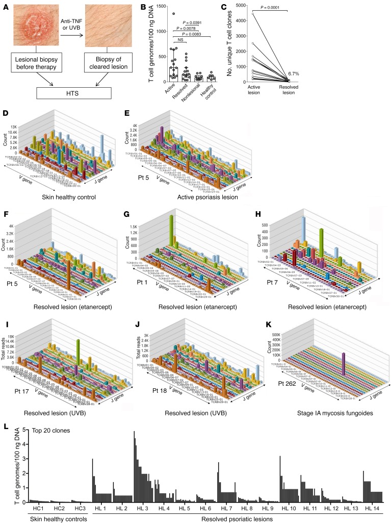
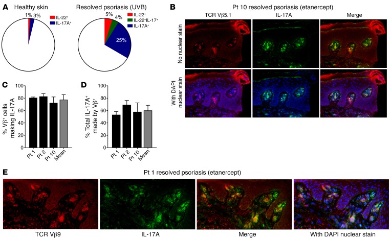
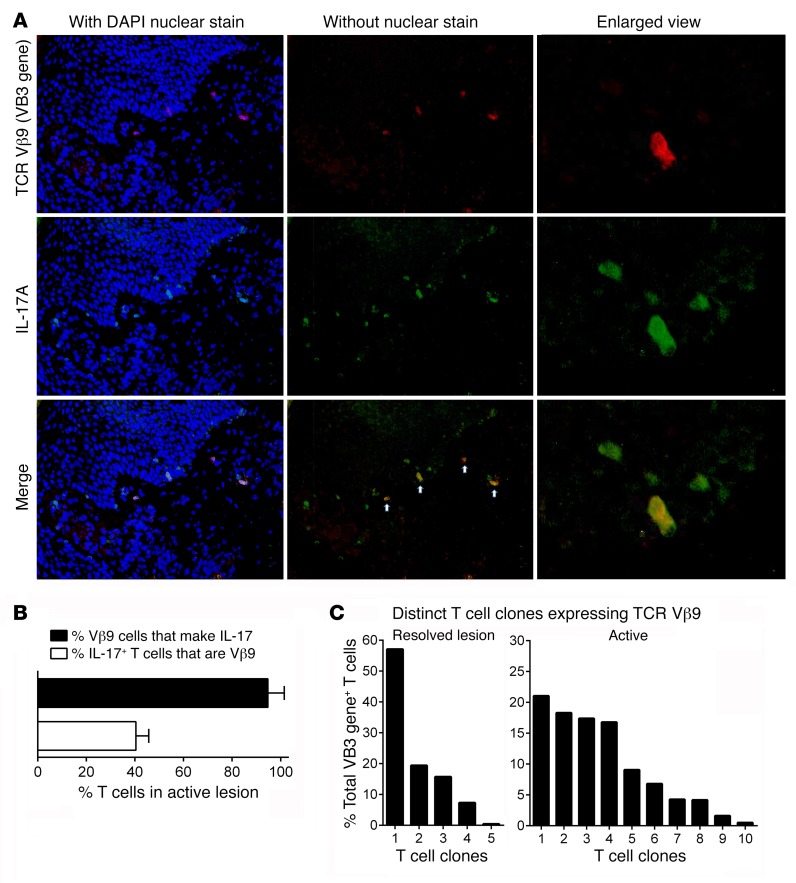
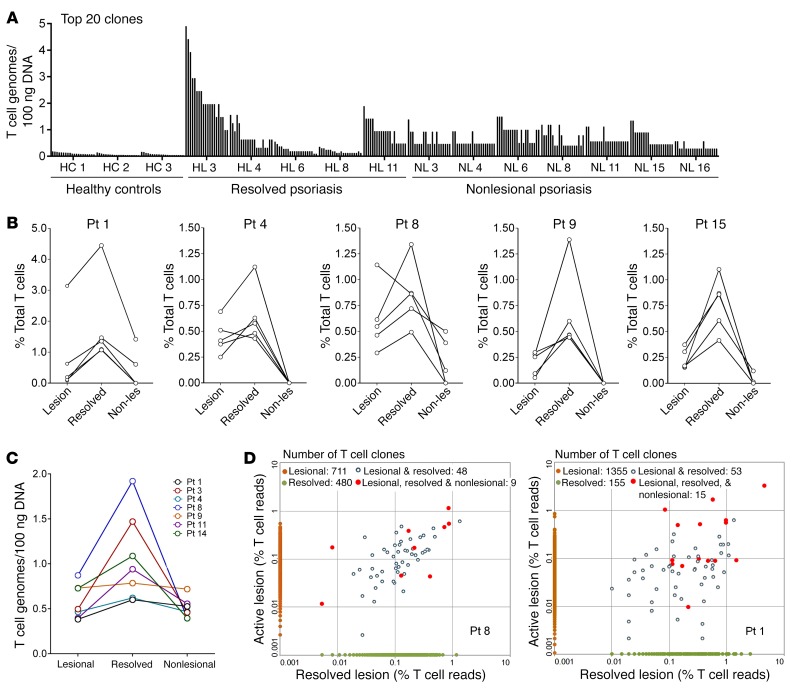
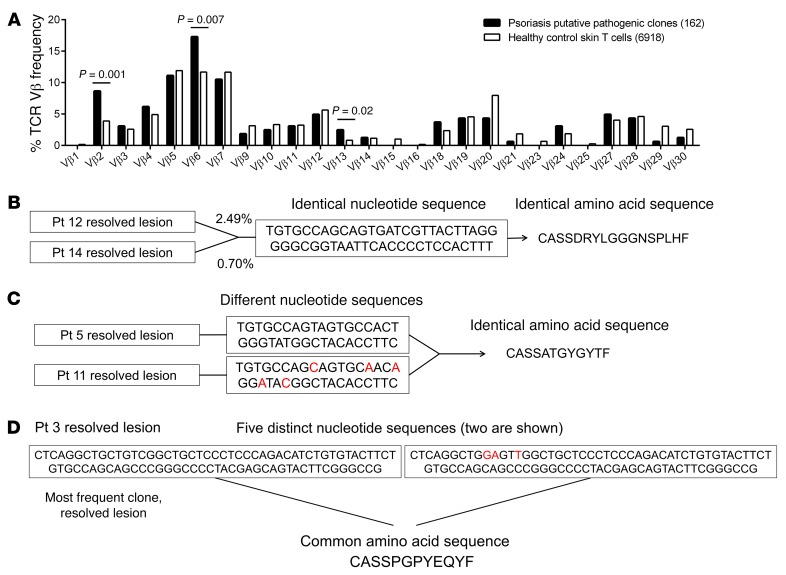
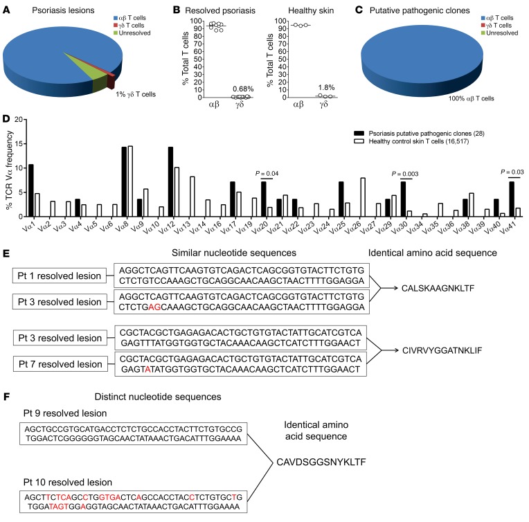
In psoriasis, an IL-17-mediated inflammatory skin disease, skin lesions resolve with therapy, but often recur in the same locations when therapy is discontinued. We propose that residual T cell populations in resolved psoriatic lesions represent the pathogenic T cells of origin in this disease. Utilizing high-throughput screening (HTS) of the T cell receptor (TCR) and immunostaining, we found that clinically resolved psoriatic lesions contained oligoclonal populations of T cells that produced IL-17A in both resolved and active psoriatic lesions. Putative pathogenic clones preferentially utilized particular Vβ and Vα subfamilies. We identified 15 TCRβ and 4 TCRα antigen receptor sequences shared between psoriasis patients and not observed in healthy controls or other inflammatory skin conditions. To address the relative roles of αβ versus γδ T cells in psoriasis, we carried out TCR/δ HTS. These studies demonstrated that the majority of T cells in psoriasis and healthy skin are αβ T cells. γδ T cells made up 1% of T cells in active psoriasis, less than 1% in resolved psoriatic lesions, and less than 2% in healthy skin. All of the 70 most frequent putative pathogenic T cell clones were αβ T cells. In summary, IL-17-producing αβ T cell clones with psoriasis-specific antigen receptors exist in clinically resolved psoriatic skin lesions. These cells likely represent the disease-initiating pathogenic T cells in psoriasis, suggesting that lasting control of this disease will require suppression of these resident T cell populations.


Conflict of interest statement

Conflict of interest: The authors have declared that no conflict of interest exists.

Figures

Figure 1
Figure 1. Oligoclonal populations of T cells are present in clinically resolved psoriatic skin lesions.
(A) Experimental design: single psoriatic lesions were biopsied before and after clearance on etanercept (anti-TNF) or UVB therapy, and T cells were evaluated using HTS of the TCR. (B) The total T cells per unit skin (100 ng of skin DNA) are shown for active psoriatic lesions (active, n = 15), clinically resolved lesions following etanercept therapy (resolved, n = 15), nonlesional skin (n = 10) (samples compared by Wilcoxon matched-paired signed rank test), and the skin of healthy individuals (healthy control, n = 6) (samples compared by Mann-Whitney U tests). (C) The number of unique T cell clones, as measured by the total number of unique CDR3 sequences, are shown for 14 patients before (active lesion) and after (resolved lesion) clinical resolution of psoriasis on etanercept therapy (Wilcoxon matched-paired signed rank test). The total numbers of unique T cell clones decreased by a mean of 93.3% following clinical clearance. (DK) The skin T cell repertoires of a healthy control (D), an active psoriatic lesion (E), resolved psoriatic lesions after clearance on etanercept (FH) or UVB therapy (I and J), and the clonal T cell lymphoproliferative disease mycosis fungoides (K) are shown. Oligoclonal populations of T cells were evident in resolved psoriatic lesions. Pt, patient. (L) The absolute number of individual T cell clones per unit skin (100 ng total skin DNA) of the top 20 clones are shown for 3 healthy controls and resolved psoriatic lesions from 14 etanercept-treated patients.
Figure 2
Figure 2. Residual T cell populations and expanded T cell clones in clinically resolved psoriatic lesions produce key pathogenic cytokines in psoriasis.
(A) IL-17A and IL-22 are produced by residual T cell populations in clinically resolved psoriatic lesions. T cells were isolated from biopsies of clinically resolved psoriasis after UVB phototherapy, and their cytokine production was studied by intracellular cytokine staining and flow cytometry. The mean results of healthy skin (n = 4) and resolved psoriasis lesions (n = 3) are shown. (BE) Expanded T cell clones identified by HTS produce IL-17A in healed lesions. HTS was used to identify the TCR Vβ subunit used by T cell clones expanded in resolved lesions. Three patients in the cohort had oligoclones in resolved lesions that used the VB03 or VB05 genes that correspond to the use of the TCR Vβ9 and Vβ5.1 protein subunits by T cells, respectively. Costaining for (B) Vβ5.1-expressing and (E) Vβ9-expressing T cells and IL-17A in these patients is shown. Individual values and the mean from 3 patients of the (C) percentage of Vβ+ T cells producing IL-17A and (D) the proportion of total IL-17A produced by Vβ+ T cells are shown.
Figure 3
Figure 3. T cell oligoclones identified in resolved lesions also produce IL-17A in active lesions from the same patient.
Studies of the active psoriatic lesion from patient 15 are shown. This patient had expanded VB03 gene/Vβ9 protein–expressing T cells in resolved lesions. (A) Costaining for Vβ9-expressing T cells and IL-17A are shown. (B) Over 90% of Vβ9 T cells produced IL-17A in active lesions, and Vβ9-expressing T cells contributed approximately 40% of the T cell–derived IL-17A in the active lesion. (C) However, this population of Vβ9 T cells contained 5 T cell clones in the resolved lesion (one of which made up 57% of the total Vβ9 population), but the active lesion from the same patient had 10 distinct Vβ9 T cell clones contributing to this population. Total Vβ9 T cell–derived IL-17A therefore contains contributions from Vβ9 T cell clones recruited into skin in the active lesion and is not an exact measurement of the contribution of putative pathogenic V9 T cell clones to total IL-17A production. Results from patient 15 are shown; comparable results were obtained in 2 additional patients.
Figure 4
Figure 4. Putative pathogenic T cell clones are also present in lower numbers in the nonlesional skin from psoriasis patients.
(A) Expanded oligoclonal populations of T cells are present in nonlesional skin. The total T cells per unit skin (100 ng of skin DNA) of the top 20 most frequent T cell clones are shown for nonlesional skin from 7 psoriatic patients (nonlesional psoriasis [NL], far right). Three skin samples from healthy individuals (healthy controls [HC]) and 4 clinically resolved psoriatic lesions (resolved psoriasis, healed lesion [HL]) are included for comparison. (B) Putative pathogenic T cells are detected in the nonlesional skin of patients with psoriasis. Putative pathogenic clones were defined as the most frequent T cell clones in resolved lesions that were also present in active lesions. Clone-tracking analyses were used to measure the frequency of the top 5 most frequent of these T cell clones in active lesions (Lesion), resolved lesions (Resolved), and nonlesional skin from the same patient (Non-les). The frequency of these T cell clones was highest in resolved lesions, followed by active lesions, and was lowest in the nonlesional skin. Data from 5 patients are shown; 5 additional patients showed a similar pattern. (C) In addition to being more frequent, putative pathogenic clones were present in highest absolute numbers in resolved lesions. The absolute number of T cells per 100 ng of total DNA is shown for the most frequent putative pathogenic T cell clone in 7 patients. (D) Overlap analyses demonstrate that shared T cell clones among lesional, resolved, and nonlesional skin (red circles) are among the most frequent T cell clones in both active and resolved psoriatic lesions. Two representative analyses out of a total of 7 are shown.
Figure 5
Figure 5. Putative pathogenic T cell clones utilized a skewed TCR Vβ repertoire and had unique TCRs not shared with healthy controls.
(A) The TCR Vβ gene usage of putative pathogenic T cell clones from 14 psoriasis patients was compared with that in a pool of 6,918 T cell CDR3 sequences from the skin of 6 healthy controls. Use of TCR Vβ2, V6, and Vβ13 was significantly more frequent among putative pathogenic T cell clones (χ2 test). (BD) T cells from different psoriatic patients had identical TCRβ CDR3 amino acid sequences that were not observed in healthy controls. (B) In 8 psoriasis patients, T cells with identical CDR3 nucleotide sequences giving rise to identical amino acid sequences were found in 2 different patients; these sequences were not observed in healthy controls. The sequences of all shared receptors are also shown in Table 1. (C) In 11 cases, different CDR3 nucleotide sequences in 2 different patients converged to produce the same amino acid sequence; these sequences were not observed in healthy controls. (D) Within individual psoriasis patients, multiple distinct nucleotide sequences were found in 7 patients who produced the same amino acid sequence; these sequences were also not observed in healthy controls.
Figure 6
Figure 6. αβ T cells predominate in psoriatic and healthy human skin, and putative pathogenic clones in psoriasis were αβ T cells that utilized a skewed Vα repertoire and contained common TCRα CDR3 sequences not observed in healthy controls.
(AC) Sequencing of the TCRα/δ locus revealed that αβ T cells predominate in psoriatic lesions and healthy human skin and putative pathogenic T cells were universally α T cells. The percentages of αβ versus γδ T cells in (A) active psoriatic lesions (n = 10), (B) resolved psoriatic lesions (n = 10), and skin from healthy controls (n = 3), and among (C) putative pathogenic T cell clones in psoriasis are shown. Results are also summarized in Table 2. (D) Putative pathogenic T cell clones preferentially utilized Vα20, Vα30, and Vα41, as compared with 16,517 TCRα sequences from the skin of 6 healthy controls (χ2 test). (E and F) T cells with highly similar TCR Vα CDR3 sequences (1 or 2 nucleotides different, 4 pairs of patients) or clearly distinct Vα CDR3 sequences (1 patient pair) giving rise to identical CDR3 amino acid sequences were found in common between patients with psoriasis. These shared CDR3 amino acid sequences were not observed in healthy controls. Shared receptors are listed in Table 1.

Similar articles

Cited by

References

    1. Krueger JG, et al. IL-17A is essential for cell activation and inflammatory gene circuits in subjects with psoriasis. J Allergy Clin Immunol. 2012;130(1):145–154.e9. doi: 10.1016/j.jaci.2012.04.024. - DOI - PMC - PubMed
    1. Golden JB, McCormick TS, Ward NL. IL-17 in psoriasis: implications for therapy and cardiovascular co-morbidities. Cytokine. 2013;62(2):195–201. doi: 10.1016/j.cyto.2013.03.013. - DOI - PMC - PubMed
    1. Suárez-Fariñas M, Fuentes-Duculan J, Lowes MA, Krueger JG. Resolved psoriasis lesions retain expression of a subset of disease-related genes. J Invest Dermatol. 2011;131(2):391–400. doi: 10.1038/jid.2010.280. - DOI - PMC - PubMed
    1. Cheuk S, et al. Epidermal Th22 and Tc17 cells form a localized disease memory in clinically healed psoriasis. J Immunol. 2014;192(7):3111–3120. doi: 10.4049/jimmunol.1302313. - DOI - PMC - PubMed
    1. Boyman O, Hefti HP, Conrad C, Nickoloff BJ, Suter M, Nestle FO. Spontaneous development of psoriasis in a new animal model shows an essential role for resident T cells and tumor necrosis factor-alpha. J Exp Med. 2004;199(5):731–736. doi: 10.1084/jem.20031482. - DOI - PMC - PubMed