. 2018 Jan 18;37(3):341-351.
doi: 10.1038/onc.2017.326. Epub 2017 Sep 25.

PTEN deficiency sensitizes endometrioid endometrial cancer to compound PARP-PI3K inhibition but not PARP inhibition as monotherapy

Affiliations

PTEN deficiency sensitizes endometrioid endometrial cancer to compound PARP-PI3K inhibition but not PARP inhibition as monotherapy

X Bian et al. Oncogene. .

Abstract

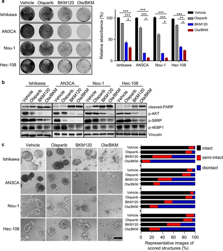
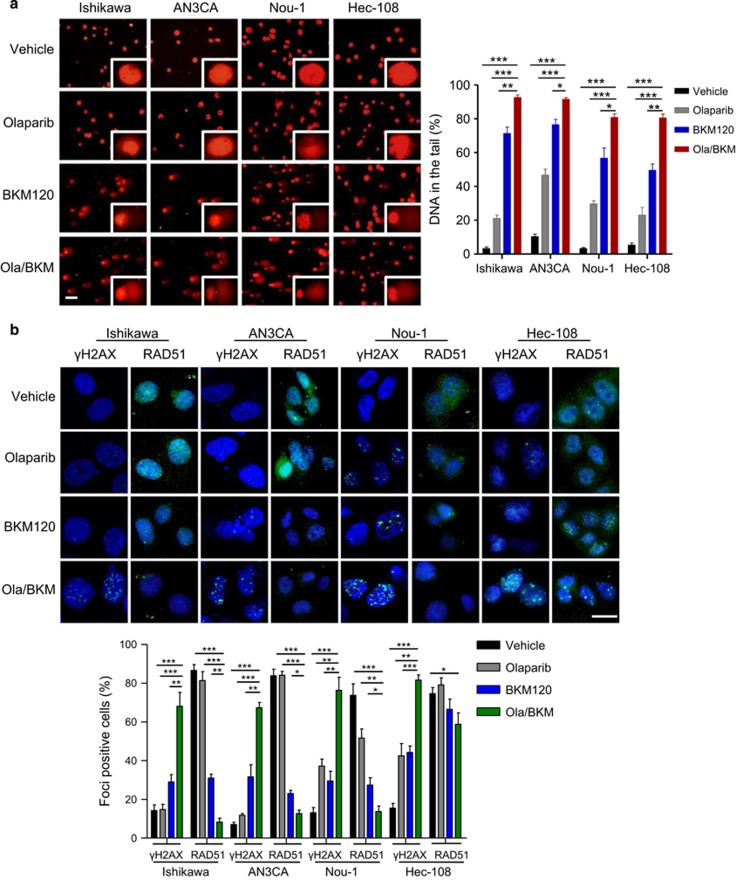
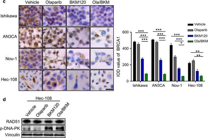
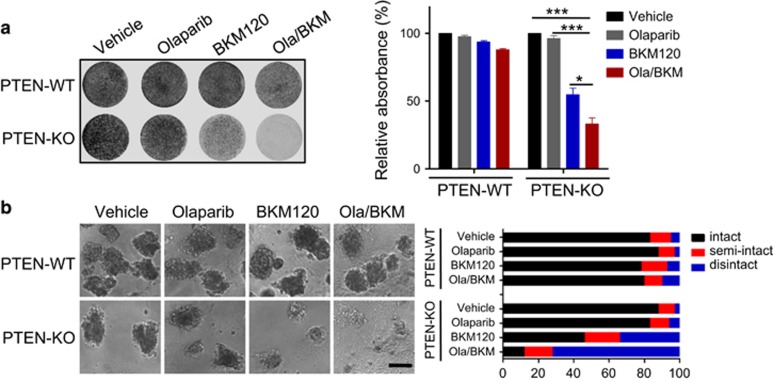
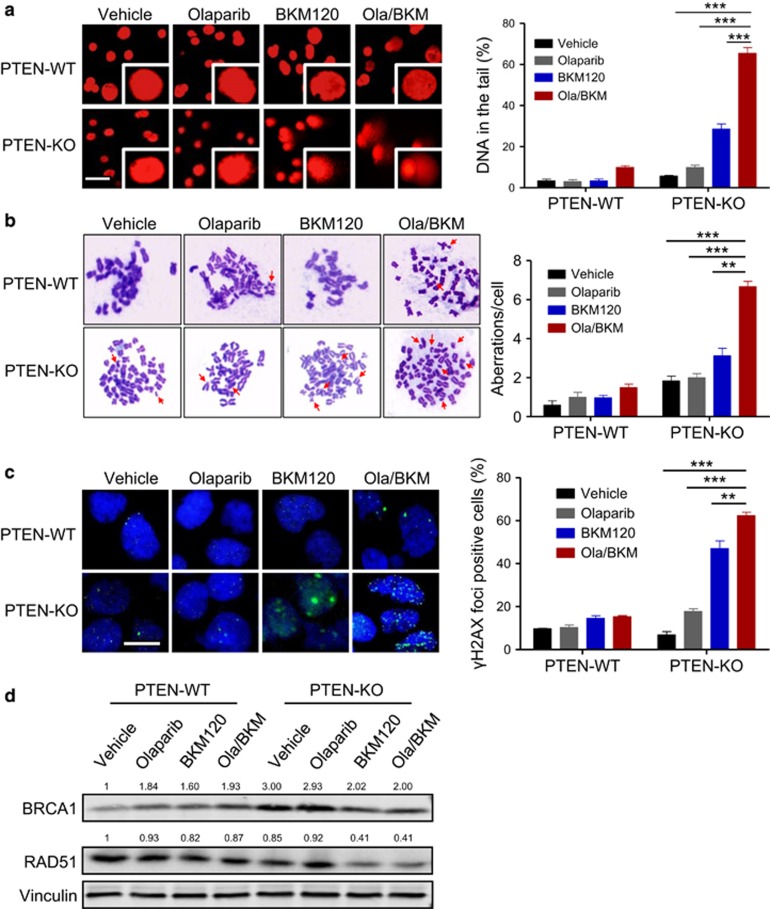
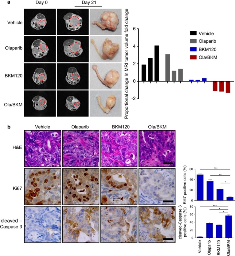
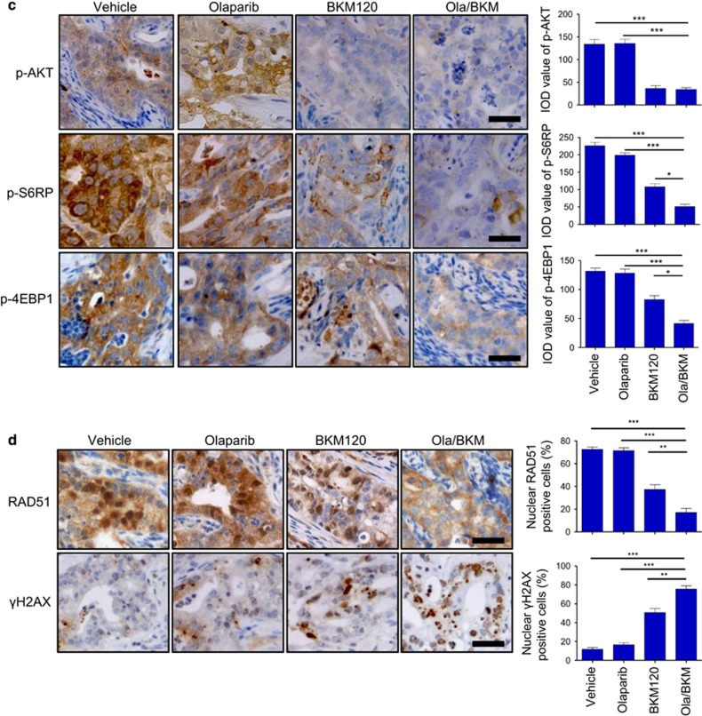
Poly (ADP-ribose) polymerase (PARP) inhibitors have emerged as promising cancer therapeutics especially for tumors with deficient homologous recombination (HR) repair. However, as HR-deficient tumors represent only a small fraction of endometrial cancers, the therapeutic utility of PARP inhibitors is limited in this disease. Somatic loss of phosphatase and tensin homolog (PTEN), a tumor suppressor that counteracts phosphoinositide 3-kinase (PI3K) activity, is one of the most common genetic aberrations in endometrioid endometrial cancer. While previous works have identified the role of PTEN in DNA double-strand break repair, vulnerabilities of PTEN-deficient endometrioid endometrial cancers to PARP inhibition remain controversial. Here we find that PTEN-deficient endometrioid endometrial cancer cells are not responsive to PARP inhibitor Olaparib alone, but instead show superior sensitivity to compound inhibition with PI3K inhibitor BKM120, as evidenced by reduced clonogenic cell growth and three-dimensional (3D) spheroid disintegration. Mechanistically, PI3K blockade by BKM120 attenuated HR competency with γH2AX accumulation and reduced RAD51 and BRCA1 expression in Ishikawa, AN3CA and Nou-1 cells, but the same combination treatment led to enhanced phosphorylation of DNA-PK, a non-homologous end joining repair protein, in Hec-108 cells. Furthermore, we show that CRISPR/Cas9-mediated PTEN depletion rendered PTEN wild-type Hec-1A endometrioid endometrial cancer cells responsive to combined inhibition of PARP/PI3K, with concomitantly induced DNA damage accumulation and repair defects. The combination of BKM120 and Olaparib cooperated to inhibit tumor growth in a genetic mouse model of Pten-deficient endometrioid endometrial cancer. Together, these results suggest PI3K inhibition may be a plausible approach to expand the utility of PARP inhibitors to endometrioid endometrial cancers in a PTEN-deficient setting.


Conflict of interest statement

The authors declare no conflict of interest.

Figures

Figure 1
Figure 1
Effects of Olaparib and BKM120 as single-agents or in combination on the survival and growth of PTEN-deficient endometrioid endometrial cancer cells. (a) PTEN-deficient endometrioid ensdometrial cancer cell lines as indicated were treated with drugs for 7–10 days. Fresh media with drugs were replaced every 3 days. At the end point, plates were fixed and stained with crystal violet. All experiments were performed in triplicate. Representative images of plates are shown. Error bars represent standard deviations (s.d.) from the mean. *P<0.05; **P<0.01; ***P<0.001 (Student’s t-test). (b) PTEN-deficient endometrioid endometrial cancer cell lines were treated with drugs as indicated for 24 h. Phosphorylated AKT, S6RP and 4EBP1 proteins and cleaved PARP were detected by western blot. Vinculin served as a loading control. (c) PTEN-deficient endometrioid endometrial cancer cell lines were cultured in 3D matrigel and drug treated for 10–12 days. Representative images of cells are shown in the left panel. Quantification of scored structures (intact, semi-disintegrated and disintegrated) is shown in the right panel. Scale bar, 100 μm.
Figure 2
Figure 2
Effects of Olaparib and BKM120 as single-agents or in combination on DNA damage and repairs in PTEN-deficient endometrioid endometrial cancer cells. (a) DNA damage was measured by comet assay in PTEN-deficient endometrioid endometrial cancer cells treated with drugs as indicated for 48 h. Scale bar, 50 μm. Quantification of DNA in the tail from three independent experiments is shown as mean±s.d. (b) Immunofluorescent staining of RAD51, γH2AX and DAPI in respective PTEN-deficient endometrioid endometrial cancer cells treated with drugs as indicated for 48 h. Scale bar, 20 μm. Cells containing more than five foci were scored as positive. Means±s.d. for three independent experiments are shown. (c) Representative images of immunocytochemical staining analyses of BRCA1 protein in PTEN-deficient endometrioid endometrial cancer cells treated with drugs as indicated for 48 h. Scale bar, 50 μm. Quantification of IOD (integrated optical density) value of BRCA1 from three independent experiments is shown as mean±s.d. (d) Western blot analysis of phosphorylated-DNA-PK in Hec-108 endometrioid endometrial cancer cells treated as indicated for 24 h. Vinculin served as a loading control. *P<0.05; **P<0.01; ***P<0.001 (Student’s t-test).
Figure 2
Figure 2
Effects of Olaparib and BKM120 as single-agents or in combination on DNA damage and repairs in PTEN-deficient endometrioid endometrial cancer cells. (a) DNA damage was measured by comet assay in PTEN-deficient endometrioid endometrial cancer cells treated with drugs as indicated for 48 h. Scale bar, 50 μm. Quantification of DNA in the tail from three independent experiments is shown as mean±s.d. (b) Immunofluorescent staining of RAD51, γH2AX and DAPI in respective PTEN-deficient endometrioid endometrial cancer cells treated with drugs as indicated for 48 h. Scale bar, 20 μm. Cells containing more than five foci were scored as positive. Means±s.d. for three independent experiments are shown. (c) Representative images of immunocytochemical staining analyses of BRCA1 protein in PTEN-deficient endometrioid endometrial cancer cells treated with drugs as indicated for 48 h. Scale bar, 50 μm. Quantification of IOD (integrated optical density) value of BRCA1 from three independent experiments is shown as mean±s.d. (d) Western blot analysis of phosphorylated-DNA-PK in Hec-108 endometrioid endometrial cancer cells treated as indicated for 24 h. Vinculin served as a loading control. *P<0.05; **P<0.01; ***P<0.001 (Student’s t-test).
Figure 3
Figure 3
Effects of PTEN loss on sensitivity to the combined Olaparib and BKM120 treatment in Hec-1A endometrioid endometrial cancer cells. (a) PTEN-proficient (PTEN-WT) and deficient (PTEN-KO #3) Hec-1A endometrioid endometrial cancer cells were treated with BKM120 and Olaparib as single-agents or in combination for 10 days. Fresh media with drugs were replaced every 3 days. At the end point, plates were fixed and stained with crystal violet. All experiments were performed in triplicate. Representative images of plates are shown. Error bars represent mean±s.d. (b) Hec-1A cells (PTEN-WT vs PTEN-KO #3) were cultured in 3D and treated with drugs as indicated for 10 days. Representative images of cells are shown in the left panel. Quantification of scored structures (intact, semi-disintegrated and disintegrated) is shown in the right panel. Scale bar, 100 μm. *P<0.05; ***P<0.001 (Student’s t-test).
Figure 4
Figure 4
Effects of PTEN loss on DNA damage and repairs in response to the combined Olaparib and BKM120 treatment in Hec-1A endometrioid endometrial cancer cells. (a) DNA damage was measured by comet assay in Hec-1A cells (PTEN-WT vs PTEN-KO #3) treated with drugs as indicated for 48 h. Scale bar, 50 μm. Quantification of DNA in the tail from three independent experiments is shown as mean±s.d. (b) Metaphase spread analysis of chromosome aberrations in Hec1-A cells (PTEN-WT vs PTEN-KO #3) after drug treatments as indicated for 48 h. Representative metaphase spreads are shown. Arrows indicate chromosomal aberrations. Mean±s.d. for three independent experiments are shown. (c) Representative images of immunofluorescent staining of γH2AX and DAPI in Hec1-A cells (PTEN-WT vs PTEN-KO #3) treated with drugs as indicated for 48 h. Scale bar, 20 μm. Cells containing more than five foci were scored as positive. Means±s.d. for three independent experiments are shown. (d) Western blot analysis of RAD51 and BRCA1 proteins in Hec1-A cells (PTEN-WT vs PTEN-KO #3) treated as indicated for 24 h. Vinculin served as a loading control. **P<0.01; ***P<0.001 (Student’s t-test).
Figure 5
Figure 5
Effects of Olaparib and BKM120 as single-agents or in combination on the genetic mouse model of endometrioid endometrial tumors driven by co-loss of Pten and Lkb1. (a) Female Ptenloxp/loxp/Lkb1loxp/loxp mice were injected with adenovirus expressing Cre recombinase (Ade-Cre). Six weeks post injection, injected mice were treated with Olaparib (50 mg/kg/day, intraperitoneal injection), BKM120 (30 mg/kg/day, oral gavage) as single-agents or in combination. Representative MRI images of mice at initiation (T0) and completion of drug treatment (21 days, T21) (left panel) and the waterfall plot depicting proportional changes in tumor volume (right panel) are shown (n=3 per treatment group). Representative images of histological (b) and immunohistochemical staining (bd) for proteins as indicated in tumors from Ade-Cre-injected Ptenloxp/loxp/Lkb1loxp/loxp mice (n=6 per treatment group) treated with BKM120 and Olaparib as single-agents or in combination for 3 days. Scale bar, 25 μm. Data are shown as mean±s.e.m. *P<0.05; **P<0.01; ***P<0.001 (Student’s t-test). H&E, hematoxylin and eosin.
Figure 5
Figure 5
Effects of Olaparib and BKM120 as single-agents or in combination on the genetic mouse model of endometrioid endometrial tumors driven by co-loss of Pten and Lkb1. (a) Female Ptenloxp/loxp/Lkb1loxp/loxp mice were injected with adenovirus expressing Cre recombinase (Ade-Cre). Six weeks post injection, injected mice were treated with Olaparib (50 mg/kg/day, intraperitoneal injection), BKM120 (30 mg/kg/day, oral gavage) as single-agents or in combination. Representative MRI images of mice at initiation (T0) and completion of drug treatment (21 days, T21) (left panel) and the waterfall plot depicting proportional changes in tumor volume (right panel) are shown (n=3 per treatment group). Representative images of histological (b) and immunohistochemical staining (bd) for proteins as indicated in tumors from Ade-Cre-injected Ptenloxp/loxp/Lkb1loxp/loxp mice (n=6 per treatment group) treated with BKM120 and Olaparib as single-agents or in combination for 3 days. Scale bar, 25 μm. Data are shown as mean±s.e.m. *P<0.05; **P<0.01; ***P<0.001 (Student’s t-test). H&E, hematoxylin and eosin.

References

    1. Amant F, Moerman P, Neven P, Timmerman D, Van Limbergen E, Vergote I. Endometrial cancer. Lancet 2005; 366: 491–505. - PubMed
    1. Cancer Genome Atlas Research NCancer Genome Atlas Research NKandoth C Cancer Genome Atlas Research NSchultz N Cancer Genome Atlas Research NCherniack AD Cancer Genome Atlas Research NAkbani R Cancer Genome Atlas Research NLiu Y et al. Integrated genomic characterization of endometrial carcinoma. Nature 2013; 497: 67–73. - PMC - PubMed
    1. Dedes KJ, Wetterskog D, Ashworth A, Kaye SB, Reis-Filho JS. Emerging therapeutic targets in endometrial cancer. Nat Rev Clin Oncol 2011; 8: 261–271. - PubMed
    1. Kim G, Ison G, McKee AE, Zhang H, Tang S, Gwise T et al. FDA approval summary: olaparib monotherapy in patients with deleterious germline BRCA-mutated advanced ovarian cancer treated with three or more lines of chemotherapy. Clin Cancer Res 2015; 21: 4257–4261. - PubMed
    1. Konstantinopoulos PA, Ceccaldi R, Shapiro GI, D'Andrea AD. Homologous recombination deficiency: exploiting the fundamental vulnerability of ovarian cancer. Cancer Discov 2015; 5: 1137–1154. - PMC - PubMed

Publication types

MeSH terms