. 2017 Nov;24(11):893-901.
doi: 10.1038/nsmb.3470. Epub 2017 Sep 25.

Atomic view of the energy landscape in the allosteric regulation of Abl kinase

Affiliations

Atomic view of the energy landscape in the allosteric regulation of Abl kinase

Tamjeed Saleh et al. Nat Struct Mol Biol. 2017 Nov.

Abstract

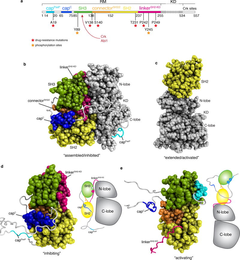
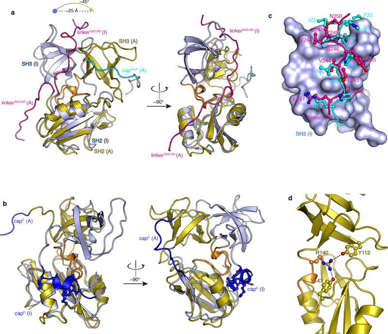
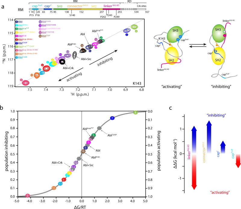
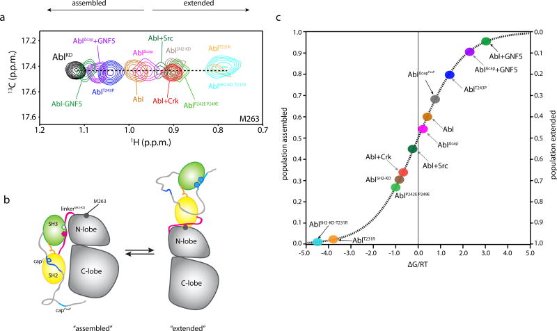
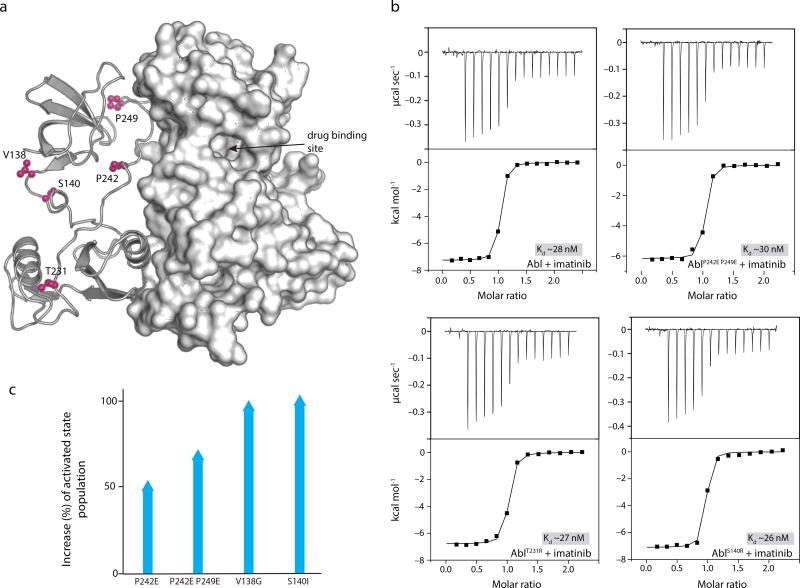
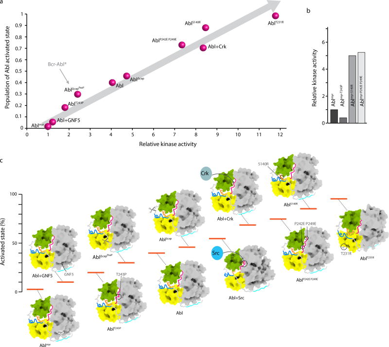
The activity of protein kinases is often regulated in an intramolecular fashion by signaling domains, which feature several phosphorylation or protein-docking sites. How kinases integrate such distinct binding and signaling events to regulate their activities is unclear, especially in quantitative terms. We used NMR spectroscopy to show how structural elements within the Abl regulatory module (RM) synergistically generate a multilayered allosteric mechanism that enables Abl kinase to function as a finely tuned switch. We dissected the structure and energetics of the regulatory mechanism to precisely measure the effects of various activating or inhibiting stimuli on Abl kinase activity. The data provide a mechanistic basis explaining genetic observations and reveal a previously unknown activator region within Abl. Our findings show that drug-resistance mutations in the Abl RM exert their allosteric effect by promoting the activated state of Abl and not by decreasing the drug affinity for the kinase.


Conflict of interest statement

COMPETING FINANCIAL INTERESTS

The authors declare no competing financial interests.

Figures

Figure 1
Figure 1
Structures of Abl. (a) Domain organization of the first N-terminal 557 residues of Abl. The regulatory module (RM) consists of the first 255 residues and its domains and motifs are indicated. The kinase (catalytic) domain (KD) encompasses residues 255–534, whereas the region following (residues 534–557) is disordered and includes a binding site for Crk. Position of imatinib-resistant mutation sites are denoted by red asterisks. Tyrosine phosphorylation sites by Hck and Src kinases are denoted by orange asterisks. CrkSH2 and Abi1 binding sites are indicated by a red arrow. (b) Crystal structure (PDB ID 2FO0) of the myristoylated form of Abl in the assembled state. The first ~56 N-terminal residues were not visible in the crystal structure and were modeled here as a disordered segment (grey). The various domains and motifs are colored per the color code in panel a. Myr indicates the myristate moiety. (c) Crystal structure (PDB ID 4XEY) of the Abl (SH2-KD) in the extended state. (d) The lowest-energy solution structure of the isolated Abl RM in its inhibiting state is shown as a space-filling model for the structured regions and as ribbon for the disordered regions. A cartoon of Abl is shown on the right indicating that the isolated Abl RM in its inhibiting state is compatible with the formation of the assembled state of Abl. (e) The lowest-energy solution structure of the isolated Abl RM in its activating state is shown as a space-filling model for the structured regions and as ribbon for the disordered regions. A cartoon of Abl is shown on the right indicating that the isolated Abl RM in its inhibiting state is compatible with the formation of the extended state of Abl. The structures of the two states are compared in Figure 2.
Figure 2
Figure 2
Structure analysis of the Abl RM activating and inhibiting states. (a) Superposition of the Abl RM activating (A) and inhibiting (I) states highlighting the orientation difference of the SH3-SH2 domains and the conformational changes of the linkerSH2-KD and capPxxP. The two structures are superimposed onto the SH2 domain. (b) Superposition of the Abl RM activating (A) and inhibiting (I) states highlighting the conformational change in capC in the two states. CapC is disordered in the activating state whereas it forms a short helix that docks on a hydrophobic track on the SH2 domain in the inhibiting state. The four capC residues that make the contacts with SH2 are Trp67, Lys70, Leu73 and Leu74. (c) Superposition of the structure of SH3 bound to the linkerSH2-KD (inhibiting state) and bound to the capPxxP motif (activating state). The SH3 domain is shown as a solvent-accessible surface in blue, the linkerSH2-KD in red ball-and-stick and the capPxxP in cyan ball-and-stick. The first few contacts to SH3 are very similar. Lys241, Pro242 and Val244 residues of the linkerSH2-KD are substituted by Arg14, Pro15 and Leu17 in capPxxP thus forming very similar contacts with SH3. However, whereas Tyr245 side chain in linkerSH2-KD is pointing towards the solvent, the topologically equivalent Pro18 in capPxxP forms intimate contacts with SH3. The contacts between SH3 and the linkerSH2-KD residues Val247, Pro249 and Asn250 are substituted by contacts with residues Ala19, Leu20 and Ile23 in capPxxP. Moreover, His21 and Phe22 provide additional contacts to SH3. (d) Structural basis for the stabilization of the activating state by the S140R mutation. Arg140 forms a bifurcated H-bond with Tyr112 (in the SH3) and Tyr147 (in the SH2) in the activating state.
Figure 3
Figure 3
Populations and energetics of Abl RM inhibiting and activating states. (a) Overlaid 1H-15N heteronuclear single quantum coherence (HSQC) spectra of the indicated Abl RM and Abl variants, showing the residue of K143. K143 is located in the connectorSH3/2 and displays the largest chemical shift range, thus providing the most sensitive probe for determining the populations of the two states in the variants. All of the other residues that have different chemical shifts in the two states show similar linear trend (Supplementary Fig. 3e–3f). Each Abl RM variant is denoted by a number and color. Important amino acid positions that were substituted are indicated on the top panel showing the domain organization of Abl. A schematic highlighting the structural changes in Abl RM as it transitions between the activating and the inhibiting states is shown on the right. (b) Populations of the inhibiting and activating states for the Abl RM and Abl variants determined by NMR from the data on panel A. The populations are plotted as a function of the associated free energy, ΔG/RT, where R is the gas constant, T the temperature and ΔG is given as GA-GI. 0.6 kcal mol−1 change in ΔG corresponds to a change by 1 unit in ΔG/RT at room temperature. As variants get closer to the free energy degeneracy (ΔG/RT=0) small changes in energy will result in substantial changes in the populations. (c) Energy contribution to the stability of the inhibiting and activating states by the four most important regions. Red arrows denote stabilization of the activating state whereas blue arrows denote stabilization of the inhibiting state.
Figure 4
Figure 4
Populations of Abl assembled and extended states. (a) Overlaid 1H-13C heteronuclear multiple quantum coherence (HMQC) spectra of the indicated Abl variants, showing the residue of M263. M263 is located at the interface between the SH2 and the kinase domain and provides the most sensitive probe for determining the populations of the two states in the variants. (b) Schematic highlighting the structural changes in Abl as it transitions between the assembled and the extended states. The position of M263 is shown as a grey circle. c, Populations of the assembled and extended states for Abl variants determined by NMR from the data on panel a. The populations are plotted as a function of the associated free energy, ΔG/RT, as in Figure 3b.
Figure 5
Figure 5
Mechanistic basis for imatinib resistance of RM mutations. (a) Cartoon of the crystal structure (PDB ID 2FO0) showing the location of the mutations (pink). The drug binding site is located in the KD and far away from the mutation sites. (b) Binding affinity of imatinib for Abl variants. The mutations do not reduce the affinity of imatinib for Abl. (c) Plot shows the increase of the Abl activated sate population as a result of the allosteric mutations in Abl RM.
Figure 6
Figure 6
Energy landscape of Abl allosteric regulation. (a) Plot of the activated state population of Abl, as measured by NMR and displayed in Figure 4c, against the measured kinase activity measured at 10 minute time-point shows a linear correlation. The kinase activity is expressed as relative to the activity of Ablmyr. The arrow pointing to AblΔcapPxxP indicates the expected population of the activated state in Bcr-Abl exclusively as a result of the removal of the first 45 residues in the fusion. (b) Kinase activity of Ablmyr variants indicating that further suppression or activation occurs even in the myristoylated form of Abl. (c) Energy landscape of Abl highlighting the changes in the activated state population, and thus of the kinase activity, caused by mutations, deletions, Crk binding and phosphorylation by Src. For clarity only the assembled structure is shown for all states. Uncropped gel images for a and b and quantification are shown in Supplementary Data Set 1.

Similar articles

Cited by

References

    1. Bradley WD, Koleske AJ. Regulation of cell migration and morphogenesis by Abl-family kinases: emerging mechanisms and physiological contexts. Journal of cell science. 2009;122:3441–3454. - PMC - PubMed
    1. Colicelli J. ABL tyrosine kinases: evolution of function, regulation, and specificity. Science Signaling. 2010;3:re6. - PMC - PubMed
    1. Hantschel O. Structure, regulation, signaling, and targeting of abl kinases in cancer. Genes & Cancer. 2012;3:436–446. - PMC - PubMed
    1. Greuber EK, Smith-Pearson P, Wang J, Pendergast AM. Role of ABL family kinases in cancer: from leukaemia to solid tumours. Nature reviews Cancer. 2013;13:559–571. - PMC - PubMed
    1. Khatri A, Wang J, Pendergast AM. Multifunctional Abl kinases in health and disease. J Cell Sci. 2016;129:9–16. - PMC - PubMed

MeSH terms

Substances

LinkOut - more resources