. 2018 Feb;97(2):192-200.
doi: 10.1177/0022034517732754. Epub 2017 Sep 25.

Complex Intratissue Microbiota Forms Biofilms in Periodontal Lesions

Affiliations

Complex Intratissue Microbiota Forms Biofilms in Periodontal Lesions

K Baek et al. J Dent Res. 2018 Feb.

Abstract

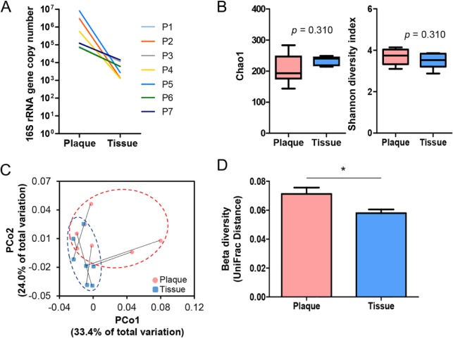
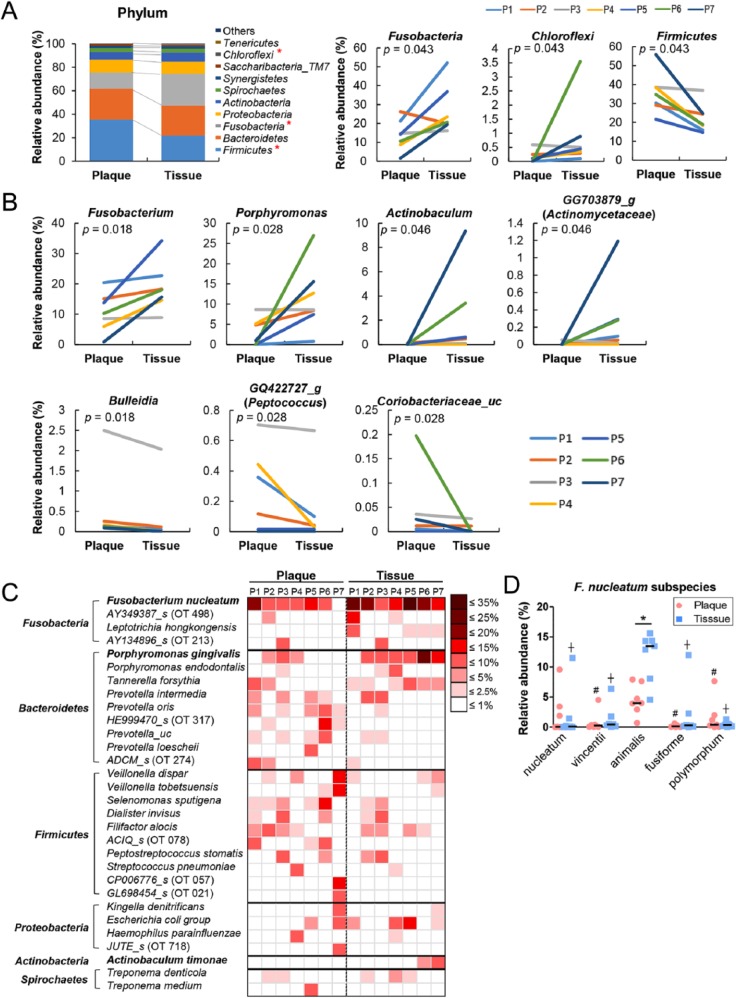
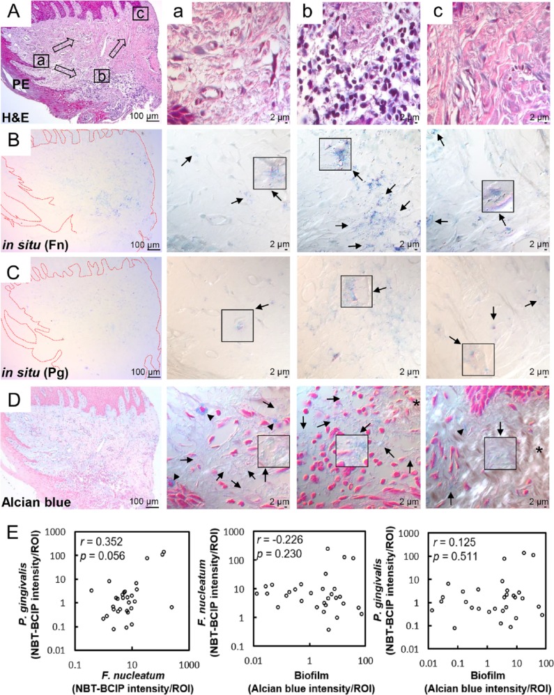
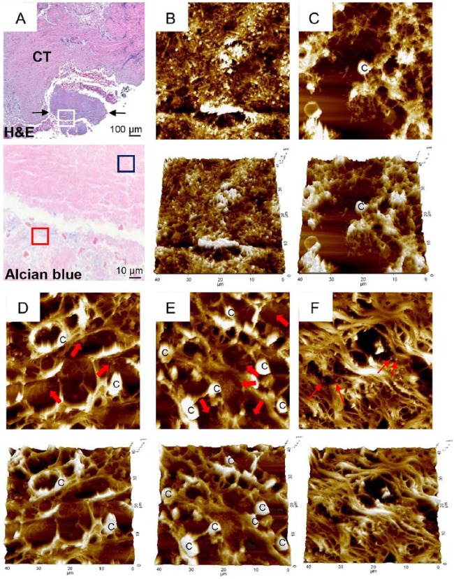
Periodontitis is caused by dysbiotic subgingival bacterial communities that may lead to increased bacterial invasion into gingival tissues. Although shifts in community structures associated with transition from health to periodontitis have been well characterized, the nature of bacteria present within the gingival tissue of periodontal lesions is not known. To characterize microbiota within tissues of periodontal lesions and compare them with plaque microbiota, gingival tissues and subgingival plaques were obtained from 7 patients with chronic periodontitis. A sequencing analysis of the 16S rRNA gene revealed that species richness and diversity were not significantly different between the 2 groups. However, intersubject variability of intratissue communities was smaller than that of plaque communities. In addition, when compared with the plaque communities, intratissue communities were characterized by decreased abundance of Firmicutes and increased abundance of Fusobacteria and Chloroflexi. In particular, Fusobacterium nucleatum and Porphyromonas gingivalis were highly enriched within the tissue, composing 15% to 40% of the total bacteria. Furthermore, biofilms, as visualized by alcian blue staining and atomic force microscopy, were observed within the tissue where the degradation of connective tissue fibers was prominent. In conclusion, very complex bacterial communities exist in the form of biofilms within the gingival tissue of periodontal lesions, which potentially serve as a reservoir for persistent infection. This novel finding may prompt new research on therapeutic strategies to treat periodontitis.

Keywords: Fusobacterium nucleatum; bacteria; gingiva; metagenomics; microbial ecology; periodontitis.


Conflict of interest statement

The authors declare no potential conflicts of interest with respect to the authorship and/or publication of this article.

Figures

Figure 1.
Figure 1.
Alpha and beta diversities of subgingival plaque and intratissue communities within periodontal lesions. Subgingival plaque and gingival tissue samples were obtained from 7 patients with chronic periodontitis. Bacterial genomic DNA was prepared as described in the Materials and Methods section and subjected to MiSeq sequencing of the 16S rRNA gene. (A) Total bacterial load in each sample was estimated by real-time polymerase chain reaction with universal primers targeting the bacterial 16S rRNA gene. The total bacterial load was expressed as the 16S rRNA gene copy number in the total DNA obtained from each sample. P, patient. (B) The Chao1 and Shannon index are expressed with box and whisker plots (P value by 2-tailed Wilcoxon signed-rank test). (C) The principal coordinates analysis (PCoA) plot was generated with weighted UniFrac metric. Samples from the same subject are connected with a solid line. (D) The intersubject UniFrac distances of the subgingival plaque and intratissue communities were obtained with a weighted metric. *P < 0.05 by 2-tailed Wilcoxon signed-rank test.
Figure 2.
Figure 2.
Differences in bacterial composition between the subgingival plaque and intratissue communities. The relative abundance of each taxon between the subgingival plaque and intratissue communities was compared. P, patient. (A) The members of top 10 phyla are shown (left panel). Three phyla were differently distributed between the 2 communities (right panel). (B) Seven genera were differently distributed between the 2 communities (P value by 2-tailed Wilcoxon signed-rank test). (C) A heat map was generated for the species/phylotypes whose relative abundance was >2.5% in any sample. (D) The relative abundance of Fusobacterium nucleatum subspecies is shown. #P < 0.05 compared with animalis subspecies in plaques. P < 0.05 compared with animalis subspecies in tissues by Kruskal-Wallis test, followed by Mann-Whitney U test. *P < 0.05 by 2-tailed Wilcoxon signed-rank test.
Figure 3.
Figure 3.
Distribution of Fusobacterium nucleatum, Porphyromonas gingivalis, and biofilm within the gingival tissue. (A) Tissue sections were stained with hematoxylin and eosin (H&E). Three areas (a, b, c) were examined under high magnification (×1,000) with differential interference contrast microscopy. Arrows indicate the potential directions of infection spread. PE, pocket epithelium. (B, C) Tissue sections were in situ hybridized with F. nucleatum– and P. gingivalis–specific probes, respectively. Arrows, bacterial signals; insets, areas with biofilm-like structure are magnified. (D) Tissue sections were stained with 1% alcian blue for acid mucopolysaccharide and counterstained with nuclear fast red. Arrows, biofilm-like structures; arrowheads, mast cells; asterisk, area with intact connective tissue fibers. (E) Correlation plots between bacterial signals and biofilm formation (r and P values by Spearman’s rank correlation test). ROI, region of interest. See Appendix Figure 3 for DIC images taken by confocal microscopy.
Figure 4.
Figure 4.
Atomic force microscopy examination of biofilm. (A) A piece of plaque biofilm coembedded with tissue was located in a hematoxylin and eosin (H&E)–stained section (between 2 arrows in top panel). CT, connective tissue. Area corresponding to the white-boxed region in the H&E-stained section was examined in the serial section stained with alcian blue under high magnification (×1,000, bottom panel). Two typical areas were chosen based on alcian blue staining and examined by atomic force microscopy: (B) blue- and (C) red-boxed areas from panel A. Areas a–c (DF, respectively) from Figure 3A were examined by atomic force microscopy. Thick arrows indicate biofilm-like structures within tissue. Thin arrows indicate scattered bacterial cells. C, eukaryotic cell.

References

    1. Abusleme L, Dupuy AK, Dutzan N, Silva N, Burleson JA, Strausbaugh LD, Gamonal J, Diaz PI. 2013. The subgingival microbiome in health and periodontitis and its relationship with community biomass and inflammation. ISME J. 7(5):1016–1025. - PMC - PubMed
    1. Baek KJ, Choi YS, Kang CK, Choi Y. 2017. The proteolytic activity of Porphyromonas gingivalis is critical in a murine model of periodontitis. J Periodontol. 88(2):218–224. - PubMed
    1. Bosshardt DD, Lang NP. 2005. The junctional epithelium: from health to disease. J Dent Res. 84(1):9–20. - PubMed
    1. Carranza FA, Jr, Saglie R, Newman MG, Valentin PL. 1983. Scanning and transmission electron microscopic study of tissue-invading microorganisms in localized juvenile periodontitis. J Periodontol. 54(10):598–617. - PubMed
    1. Cekici A, Kantarci A, Hasturk H, Van Dyke TE. 2014. Inflammatory and immune pathways in the pathogenesis of periodontal disease. Periodontol 2000. 64(1):57–80. - PMC - PubMed

Publication types

LinkOut - more resources