

Retinal thinning in amyotrophic lateral sclerosis patients without ophthalmic disease
- PMID: 28945811
- PMCID: PMC5612691
- DOI: 10.1371/journal.pone.0185242
Retinal thinning in amyotrophic lateral sclerosis patients without ophthalmic disease
Abstract
Importance: Amyotrophic lateral sclerosis (ALS) is a fatal, rapidly progressive neurodegenerative disease that primarily affects motor neurons. Recently, three causative genes have been implicated in both ALS and glaucoma. However, it is still uncertain whether patients with ALS have neurodegeneration in their retinas. If so, retinal thickness measurements might be a useful biomarker for ALS progression. Previous work in this area has been inconclusive, as it has not taken into account the effect of ophthalmic diseases on retinal thinning.
Objective: To determine whether there are differences in retinal neurons in ALS patients utilizing spectral-domain optical coherence tomography (SD-OCT). We tested the hypothesis that ALS patients exhibit retinal neurodegeneration that is not associated with ophthalmic diseases.
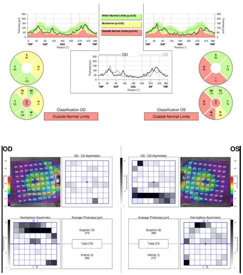
Design, settings and participants: Observational, comparative, cross-sectional study performed on patients recruited from the Duke University Medical Center ALS clinic. Patients underwent a comprehensive ophthalmologic examination to rule out ocular pathology. 21 patients met inclusion criteria. Two eyes with ocular pathology were excluded, leading to a total of 40 eyes of 21 patients included in the study. Retinal neurodegeneration was assessed by retinal nerve fiber layer (RNFL) thickness measurement using SD-OCT (Spectralis; Heidelberg Engineering).
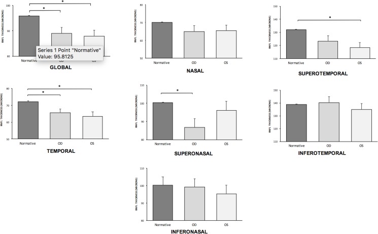
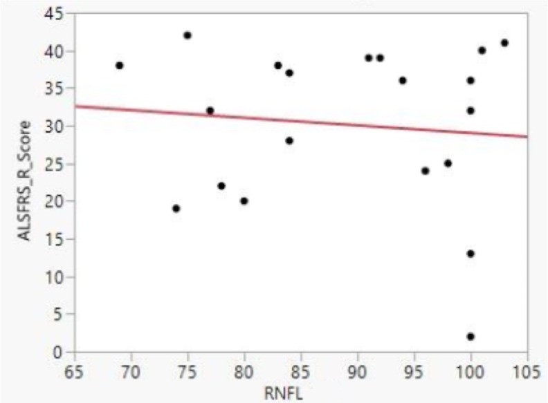
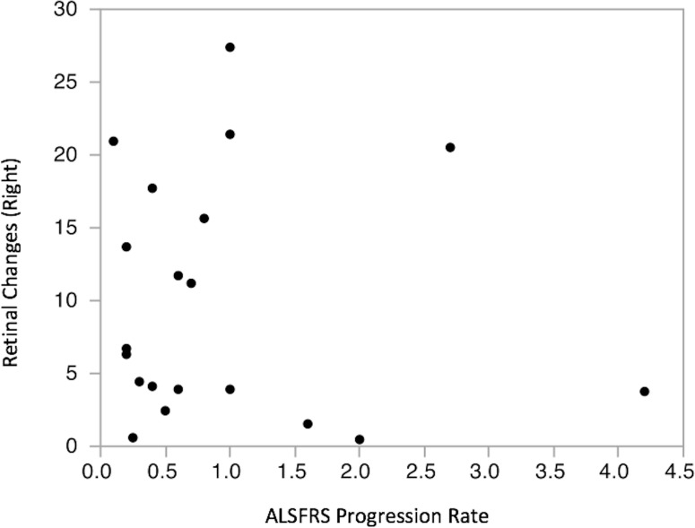
Main outcomes and measures: ALS disease severity, determined through the ALS Functional Rating Scale (ALSFRS-R); mean and six sector RNFL thickness values compared to age-adjusted values in the normative database provided by Heidelberg Engineering; RNFL thickness correlation with ALSFRS-R, ALSFRS-R progression rate, forced vital capacity (FVC), and visual acuity.
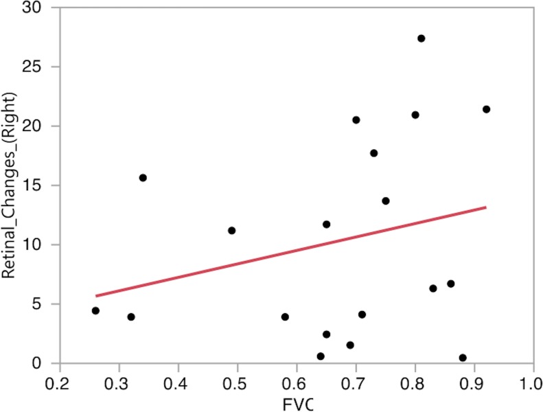
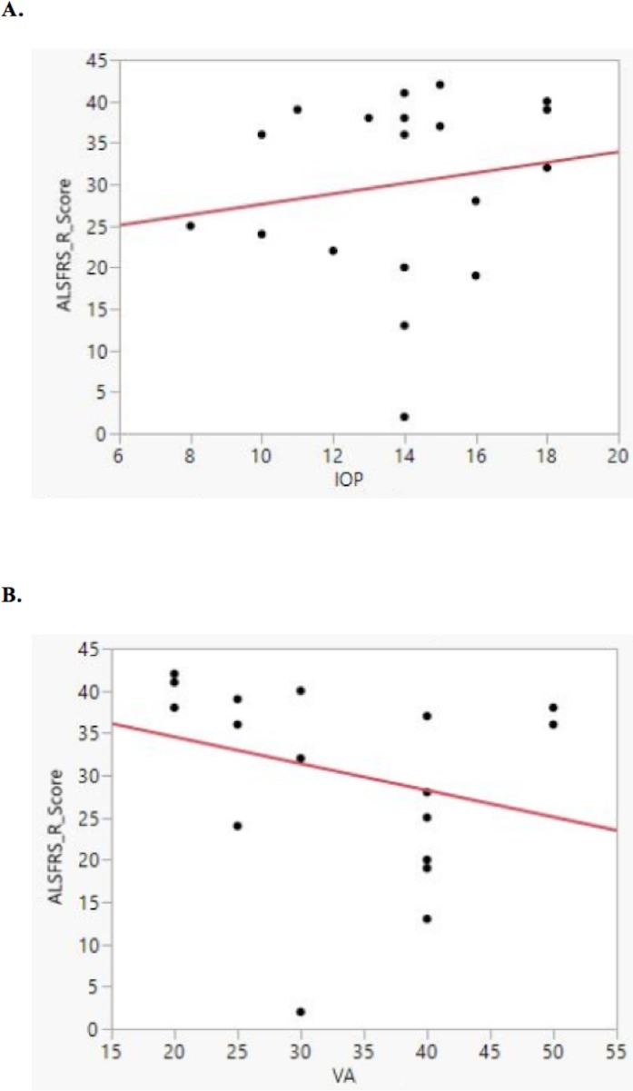
Results: ALSFRS-R mean score was 30+/-10. Mean RNFL thickness in ALS patients was 88.95 +/- 10.8 microns, significantly thinner than values in the normative database (95.81 +/- 0.8). These RNFL thickness values did not demonstrate correlation to ALSFRS-R score, ALSFRS-R progression rate, FVC, intraocular pressure, or visual acuity.
Conclusions: Using SD-OCT, our study shows that ALS patients without ocular pathology exhibit thinned retinal layers. Future studies are warranted to clarify the clinical relationship between retinal thinning and motor neuron loss in ALS.
Conflict of interest statement
Figures
References
-
- Connolly S, Galvin M, Hardiman O. End-of-life management in patients with amyotrophic lateral sclerosis. Lancet Neurol. 2015. April;14(4):435–42. doi: 10.1016/S1474-4422(14)70221-2 - DOI - PubMed
-
- Turner MR, Swash M. The expanding syndrome of amyotrophic lateral sclerosis: a clinical and molecular odyssey. J Neurol Neurosurg Psychiatry. 2015. June;86(6):667–73. doi: 10.1136/jnnp-2014-308946 - DOI - PMC - PubMed
-
- Maruyama H, Morino H, Ito H, Izumi Y, Kato H, Watanabe Y, et al. Mutations of optineurin in amyotrophic lateral sclerosis. Nature. 2010. May 13;465(7295):223–6. doi: 10.1038/nature08971 - DOI - PubMed
-
- Cirulli ET, Lasseigne BN, Petrovski S, Sapp PC, Dion PA, Leblond CS, et al. Exome sequencing in amyotrophic lateral sclerosis identifies risk genes and pathways. Science. 2015. March 27;347(6229):1436–41. doi: 10.1126/science.aaa3650 - DOI - PMC - PubMed
-
- Elden AC, Kim H, Hart MP, Chen-Plotkin AS, Johnson BS, Fang X, et al. Ataxin-2 intermediate-length polyglutamine expansions are associated with increased risk for ALS. Nature. 2010. August 26;466(7310):1069–75. doi: 10.1038/nature09320 - DOI - PMC - PubMed
Publication types
MeSH terms
LinkOut - more resources
Full Text Sources
Other Literature Sources
Medical
Miscellaneous
