

Role of proton-coupled folate transporter in pemetrexed resistance of mesothelioma: clinical evidence and new pharmacological tools
- PMID: 28945836
- PMCID: PMC5808668
- DOI: 10.1093/annonc/mdx499
Role of proton-coupled folate transporter in pemetrexed resistance of mesothelioma: clinical evidence and new pharmacological tools
Abstract
Background: Thymidylate synthase (TS) has a predictive role in pemetrexed treatment of mesothelioma; however, additional chemoresistance mechanisms are poorly understood. Here, we explored the role of the reduced-folate carrier (RFC/SLC19A1) and proton-coupled folate transporter (PCFT/SLC46A1) in antifolate resistance in mesothelioma.
Patients and methods: PCFT, RFC and TS RNA and PCFT protein levels were determined by quantitative RT-PCR of frozen tissues and immunohistochemistry of tissue-microarrays, respectively, in two cohorts of pemetrexed-treated patients. Data were analyzed by t-test, Fisher's/log-rank test and Cox proportional models. The contribution of PCFT expression and PCFT-promoter methylation to pemetrexed activity were evaluated in mesothelioma cells and spheroids, through 5-aza-2'-deoxycytidine-mediated demethylation and siRNA-knockdown.
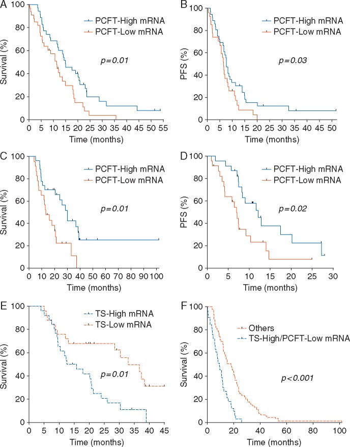
Results: Pemetrexed-treated patients with low PCFT had significantly lower rates of disease control, and shorter overall survival (OS), in both the test (N = 73, 11.3 versus 20.1 months, P = 0.01) and validation (N = 51, 12.6 versus 30.3 months, P = 0.02) cohorts. Multivariate analysis confirmed PCFT-independent prognostic role. Low-PCFT protein levels were also associated with shorter OS. Patients with both low-PCFT and high-TS levels had the worst prognosis (OS, 5.5 months), whereas associations were neither found for RFC nor in pemetrexed-untreated patients. PCFT silencing reduced pemetrexed sensitivity, whereas 5-aza-2'-deoxycytidine overcame resistance.
Conclusions: These findings identify for the first time PCFT as a novel mesothelioma prognostic biomarker, prompting prospective trials for its validation. Moreover, preclinical data suggest that targeting PCFT-promoter methylation might eradicate pemetrexed-resistant cells characterized by low-PCFT expression.
Keywords: PCFT; expression; malignant pleural mesothelioma; methylation; outcome; pemetrexed.
© The Author 2017. Published by Oxford University Press on behalf of the European Society for Medical Oncology. All rights reserved. For permissions, please email: journals.permissions@oup.com.
Figures


Similar articles
-
The promise and challenges of exploiting the proton-coupled folate transporter for selective therapeutic targeting of cancer.Cancer Chemother Pharmacol. 2018 Jan;81(1):1-15. doi: 10.1007/s00280-017-3473-8. Epub 2017 Nov 10. Cancer Chemother Pharmacol. 2018. PMID: 29127457 Free PMC article. Review.
-
Impact of hypoxia on chemoresistance of mesothelioma mediated by the proton-coupled folate transporter, and preclinical activity of new anti-LDH-A compounds.Br J Cancer. 2020 Aug;123(4):644-656. doi: 10.1038/s41416-020-0912-9. Epub 2020 Jun 4. Br J Cancer. 2020. PMID: 32493992 Free PMC article.
-
Functional Characterization of Reduced Folate Carrier and Protein-Coupled Folate Transporter for Antifolates Accumulation in Non-Small Cell Lung Cancer Cells.Drug Metab Dispos. 2024 Oct 16;52(11):1332-1344. doi: 10.1124/dmd.124.001872. Drug Metab Dispos. 2024. PMID: 39261014
-
Proton-coupled folate transporter as a biomarker of outcome to treatment for pleural mesothelioma.Pharmacogenomics. 2018 Jul 1;19(10):811-814. doi: 10.2217/pgs-2018-0071. Epub 2018 Jun 19. Pharmacogenomics. 2018. PMID: 29916298 No abstract available.
-
The major facilitative folate transporters solute carrier 19A1 and solute carrier 46A1: biology and role in antifolate chemotherapy of cancer.Drug Metab Dispos. 2014 Apr;42(4):632-49. doi: 10.1124/dmd.113.055723. Epub 2014 Jan 6. Drug Metab Dispos. 2014. PMID: 24396145 Free PMC article. Review.
Cited by
-
Tumor Targeting with Novel Pyridyl 6-Substituted Pyrrolo[2,3- d]Pyrimidine Antifolates via Cellular Uptake by Folate Receptor α and the Proton-Coupled Folate Transporter and Inhibition of De Novo Purine Nucleotide Biosynthesis.J Med Chem. 2018 Mar 8;61(5):2027-2040. doi: 10.1021/acs.jmedchem.7b01708. Epub 2018 Feb 21. J Med Chem. 2018. PMID: 29425443 Free PMC article.
-
Transporters in vitamin uptake and cellular metabolism: impacts on health and disease.Life Metab. 2025 Mar 10;4(3):loaf008. doi: 10.1093/lifemeta/loaf008. eCollection 2025 Jun. Life Metab. 2025. PMID: 40444179 Free PMC article. Review.
-
Discovery of Tumor-Targeted 6-Methyl Substituted Pemetrexed and Related Antifolates with Selective Loss of RFC Transport.ACS Med Chem Lett. 2023 Nov 15;14(12):1682-1691. doi: 10.1021/acsmedchemlett.3c00326. eCollection 2023 Dec 14. ACS Med Chem Lett. 2023. PMID: 38116433 Free PMC article.
-
Ferroptosis: Cancer Stem Cells Rely on Iron until "to Die for" It.Cells. 2021 Nov 2;10(11):2981. doi: 10.3390/cells10112981. Cells. 2021. PMID: 34831207 Free PMC article. Review.
-
Substitutions that lock and unlock the proton-coupled folate transporter (PCFT-SLC46A1) in an inward-open conformation.J Biol Chem. 2019 May 3;294(18):7245-7258. doi: 10.1074/jbc.RA118.005533. Epub 2019 Mar 11. J Biol Chem. 2019. PMID: 30858177 Free PMC article.
References
-
- Ceresoli GL, Zucali PA, Favaretto AG. et al. Phase II study of pemetrexed plus carboplatin in malignant pleural mesothelioma. J Clin Oncol 2006; 24: 1443–1448. - PubMed
-
- Righi L, Papotti MG, Ceppi P. et al. Thymidylate Synthase but not excision repair cross-complementation group-1 tumor expression predicts outcome in patients with mesothelioma treated with pemetrexed-based chemotherapy. J Clin Oncol 2010; 28: 1534–1539. - PubMed
-
- Zucali PA, Giovannetti E, Destro A. et al. Thymidylate synthase and excision repair cross-complementing group-1 as predictors of responsiveness in mesothelioma patients treated with pemetrexed/carboplatin. Clin Cancer Res 2011; 17: 2581–2590. - PubMed
MeSH terms
Substances
Grants and funding
LinkOut - more resources
Full Text Sources
Other Literature Sources
Medical
