

Hybrid capture-based genomic profiling of circulating tumor DNA from patients with estrogen receptor-positive metastatic breast cancer
- PMID: 28945887
- PMCID: PMC5834148
- DOI: 10.1093/annonc/mdx490
Hybrid capture-based genomic profiling of circulating tumor DNA from patients with estrogen receptor-positive metastatic breast cancer
Abstract
Background: Genomic changes that occur in breast cancer during the course of disease have been informed by sequencing of primary and metastatic tumor tissue. For patients with relapsed and metastatic disease, evolution of the breast cancer genome highlights the importance of using a recent sample for genomic profiling to guide clinical decision-making. Obtaining a metastatic tissue biopsy can be challenging, and analysis of circulating tumor DNA (ctDNA) from blood may provide a minimally invasive alternative.
Patients and methods: Hybrid capture-based genomic profiling was carried out on ctDNA from 254 female patients with estrogen receptor-positive breast cancer. Peripheral blood samples were submitted by clinicians in the course of routine clinical care between May 2016 and March 2017. Sequencing of 62 genes was carried out to a median unique coverage depth of 7503×. Genomic alterations (GAs) in ctDNA were evaluated and compared with matched tissue samples and genomic datasets of tissue from breast cancer.
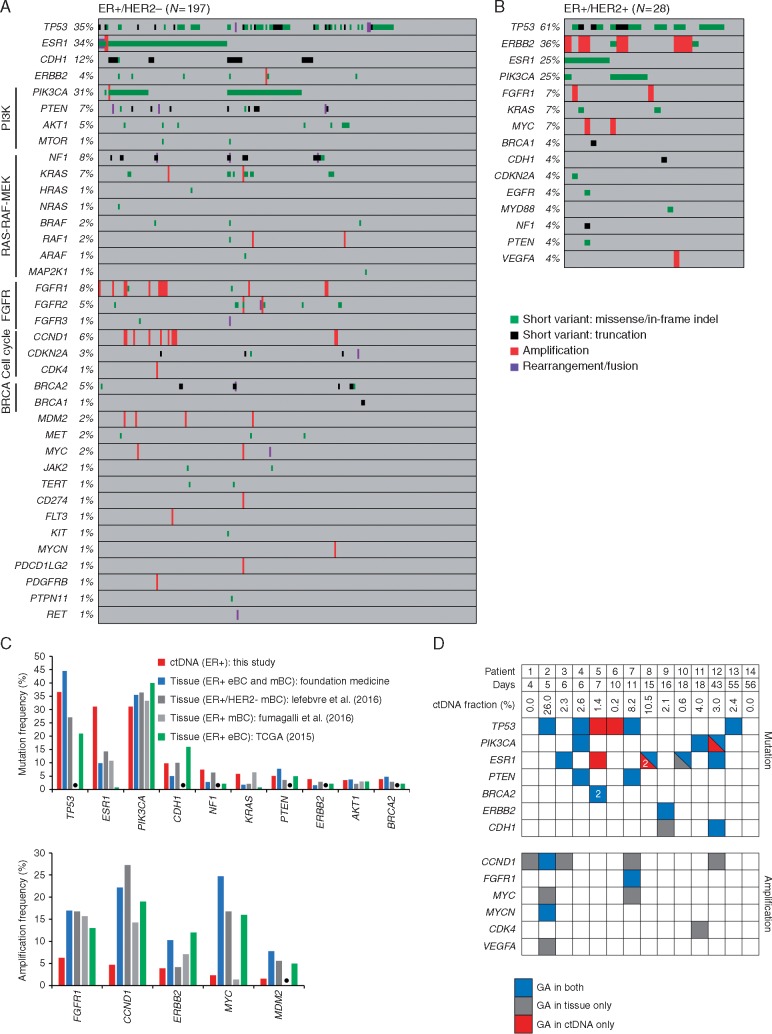
Results: At least 1 GA was reported in 78% of samples. Frequently altered genes were TP53 (38%), ESR1 (31%) and PIK3CA (31%). Temporally matched ctDNA and tissue samples were available for 14 patients; 89% of mutations detected in tissue were also detected in ctDNA. Diverse ESR1 GAs including mutation, rearrangement and amplification, were observed. Multiple concurrent ESR1 GAs were observed in 40% of ESR1-altered cases, suggesting polyclonal origin; ESR1 compound mutations were also observed in two cases. ESR1-altered cases harbored co-occurring GAs in PIK3CA (35%), FGFR1 (16%), ERBB2 (8%), BRCA1/2 (5%), and AKT1 (4%).
Conclusions: GAs relevant to relapsed/metastatic breast cancer management were identified, including diverse ESR1 GAs. Genomic profiling of ctDNA demonstrated sensitive detection of mutations found in tissue. Detection of amplifications was associated with ctDNA fraction. Genomic profiling of ctDNA may provide a complementary and possibly alternative approach to tissue-based genomic testing for patients with estrogen receptor-positive metastatic breast cancer.
Keywords: ER; ESR1; ctDNA; genomic profiling; liquid biopsy; metastatic breast cancer.
© The Author 2017. Published by Oxford University Press on behalf of the European Society for Medical Oncology.
Figures

Similar articles
-
Circulating tumour DNA in metastatic breast cancer to guide clinical trial enrolment and precision oncology: A cohort study.PLoS Med. 2020 Oct 1;17(10):e1003363. doi: 10.1371/journal.pmed.1003363. eCollection 2020 Oct. PLoS Med. 2020. PMID: 33001984 Free PMC article.
-
Clinical significance of gene mutation in ctDNA analysis for hormone receptor-positive metastatic breast cancer.Breast Cancer Res Treat. 2020 Apr;180(2):331-341. doi: 10.1007/s10549-019-05512-5. Epub 2020 Feb 4. Breast Cancer Res Treat. 2020. PMID: 32020432
-
Genomic landscape of hormone therapy-resistant HR-positive, HER2-negative breast cancer.Breast Cancer Res Treat. 2025 Sep;213(2):247-259. doi: 10.1007/s10549-025-07759-7. Epub 2025 Jul 12. Breast Cancer Res Treat. 2025. PMID: 40652157 Free PMC article.
-
ESR1 mutations in HR+/HER2-metastatic breast cancer: Enhancing the accuracy of ctDNA testing.Cancer Treat Rev. 2023 Dec;121:102642. doi: 10.1016/j.ctrv.2023.102642. Epub 2023 Oct 13. Cancer Treat Rev. 2023. PMID: 37864956 Review.
-
Standardized molecular pathology workflow for ctDNA-based ESR1 testing in HR+/HER2- metastatic breast cancer.Crit Rev Oncol Hematol. 2024 Sep;201:104427. doi: 10.1016/j.critrevonc.2024.104427. Epub 2024 Jun 23. Crit Rev Oncol Hematol. 2024. PMID: 38917944 Review.
Cited by
-
Molecular analysis of circulating tumor DNA from breast cancer patients before and after surgery and following adjuvant chemotherapy.Mol Clin Oncol. 2020 Oct;13(4):26. doi: 10.3892/mco.2020.2096. Epub 2020 Jul 20. Mol Clin Oncol. 2020. PMID: 32765873 Free PMC article.
-
Whole genome amplification of cell-free DNA enables detection of circulating tumor DNA mutations from fingerstick capillary blood.Sci Rep. 2018 Nov 23;8(1):17313. doi: 10.1038/s41598-018-35470-9. Sci Rep. 2018. PMID: 30470782 Free PMC article.
-
Liquid Biopsy to Identify Actionable Genomic Alterations.Am Soc Clin Oncol Educ Book. 2018 May 23;38:978-997. doi: 10.1200/EDBK_199765. Am Soc Clin Oncol Educ Book. 2018. PMID: 30231331 Free PMC article. Review.
-
Estrogen Receptor Alpha Mutations, Truncations, Heterodimers, and Therapies.Endocrinology. 2024 Apr 29;165(6):bqae051. doi: 10.1210/endocr/bqae051. Endocrinology. 2024. PMID: 38643482 Free PMC article. Review.
-
The Emerging Role of ESR1 Mutations in Luminal Breast Cancer as a Prognostic and Predictive Biomarker of Response to Endocrine Therapy.Cancers (Basel). 2019 Nov 28;11(12):1894. doi: 10.3390/cancers11121894. Cancers (Basel). 2019. PMID: 31795152 Free PMC article. Review.
References
-
- Fumagalli D, Wilson TR, Salgado R. et al. Somatic mutation, copy number and transcriptomic profiles of primary and matched metastatic estrogen receptor-positive breast cancers. Ann Oncol 2016; 27(10): 1860–1866. - PubMed
-
- Turner NC, Neven P, Loibl S, Andre F.. Advances in the treatment of advanced oestrogen-receptor-positive breast cancer. Lancet 2016. doi:10.1016/S0140-6736(16)32419-9. - PubMed
MeSH terms
Substances
LinkOut - more resources
Full Text Sources
Other Literature Sources
Medical
Research Materials
Miscellaneous
