Review
. 2017 Sep 25;6(10):91.
doi: 10.3390/jcm6100091.

Comparison of Minimal Residual Disease Detection by Multiparameter Flow Cytometry, ASO-qPCR, Droplet Digital PCR, and Deep Sequencing in Patients with Multiple Myeloma Who Underwent Autologous Stem Cell Transplantation

Affiliations
Review

Comparison of Minimal Residual Disease Detection by Multiparameter Flow Cytometry, ASO-qPCR, Droplet Digital PCR, and Deep Sequencing in Patients with Multiple Myeloma Who Underwent Autologous Stem Cell Transplantation

Hiroyuki Takamatsu. J Clin Med. .

Abstract

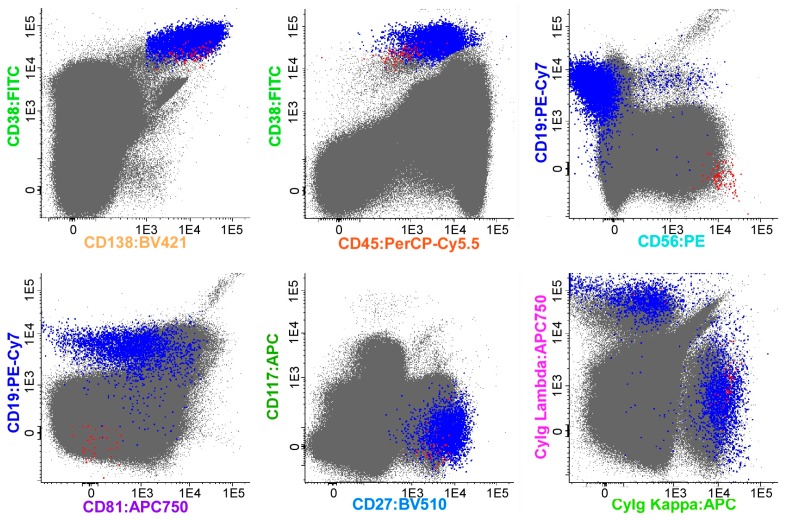
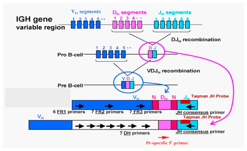
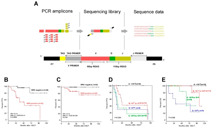
Multiple myeloma (MM) is a hematological malignancy with a poor prognosis, characterized by clonal proliferation of plasma cells in the bone marrow (BM). Relapse due to undetected minimal residual disease (MRD) is the leading cause of death among patients with MM. This review summarizes the methods and prognostic value of MRD assessment in BM and autografts from MM patients who underwent autologous stem cell transplantation (ASCT) by multiparameter flow cytometry (MFC), allele-specific oligonucleotide real-time quantitative PCR (ASO-qPCR), droplet digital PCR (ddPCR), and next-generation sequencing (NGS)-based detection methods. MRD assessment using NGS-based approaches has clear prognostic value and better sensitivity compared to traditional methods.

Keywords: allele-specific oligonucleotide-PCR; droplet digital PCR; minimal residual disease; multiple myeloma; next-generation sequencing.


Conflict of interest statement

The author received research funding from Takeda and Fujimoto, and honoraria from Celgene and Janssen.

Figures

Figure 1
Figure 1
An example of the Next-Generation Flow (NGF) method based on the analysis of (merged) data files corresponding to a bone marrow (BM) sample from multiple myeloma (MM) (>107 cells) with low levels of minimal residual disease (MRD), stained with the NGF-MM MRD panel. The figure shows bivariate dot plot representations in which plasma cells (PCs; blue and red dots) were gated using a conventional manual analysis strategy. Normal PCs (blue dots) display characteristic normal patterns of expression for the surface membrane markers used with a cytoplasmic (Cy) Igκ vs. CyIgλ ratio of 1.3. In contrast, clonal/aberrant PCs (red dots) can be clearly discriminated from normal PCs based on their more homogeneous phenotypic profile, the presence of myeloma-associated phenotypes (CD138hi, CD38dim, CD19-, CD81-, CD117-, and CD27dim), and a restricted pattern of expression of CyIgκ. Other non-PC BM populations are depicted as gray dots. Please note that, in this sample, PCs corresponded to 0.06% of all nucleated BM cells; in turn, aberrant PCs corresponded to 0.0007% of the whole BM cellularity, with an assay sensitivity (in the quantitative range) of <5 × 10⁻⁶.
Figure 2
Figure 2
Allele-specific oligonucleotide-quantitative PCR (ASO-qPCR) method to detect minimal residual disease (MRD), and design of ASO-qPCR primers and probes.
Figure 3
Figure 3
Next-generation sequencing method to detect minimal residual disease (MRD). (A) Schematic illustrations of LymphoSIGHTTM PCR primer strategy and sequencing assay. IgH V-J, IgH D-J, and IgK sequences were amplified from genomic DNA in a two-stage PCR (the first PCR using universal primer sets, and the second PCR using J primer and TAG primer). The amplified product was sequenced to obtain a high number of reads (e.g., 106 reads). The sequence reads were analyzed to determine similar sequences that form a clonotype. After clonotype determination, a standard quantitation scheme was used to calculate MRD level [43]. (B) Progression-free survival (PFS) and (C) overall survival (OS) of patients according to the MRD negativity in post-ASCT BM samples as determined by deep sequencing (threshold: 10−6). (D) PFS and (E) OS of the patients who did not receive post-ASCT treatment, according to MRD negativity in the autograft as determined by deep sequencing (threshold: 10−7).

References

    1. Attal M., Lauwers-Cances V., Marit G., Caillot D., Moreau P., Facon T., Stoppa A.M., Hulin C., Benboubker L., Garderet L., et al. Lenalidomide maintenance after stem-cell transplantation for multiple myeloma. N. Engl. J. Med. 2012;366:1782–1791. doi: 10.1056/NEJMoa1114138. - DOI - PubMed
    1. McCarthy P.L., Owzar K., Hofmeister C.C., Hurd D.D., Hassoun H., Richardson P.G., Giralt S., Stadtmauer E.A., Weisdorf D.J., Vij R., et al. Lenalidomide after stem-cell transplantation for multiple myeloma. N. Engl. J. Med. 2012;366:1770–1781. doi: 10.1056/NEJMoa1114083. - DOI - PMC - PubMed
    1. San Miguel J.F., Schlag R., Khuageva N.K., Dimopoulos M.A., Shpilberg O., Kropff M., Spicka I., Petrucci M.T., Palumbo A., Samoilova O.S., et al. Bortezomib plus melphalan and prednisone for initial treatment of multiple myeloma. N. Engl. J. Med. 2008;359:906–917. doi: 10.1056/NEJMoa0801479. - DOI - PubMed
    1. Stewart A.K., Rajkumar S.V., Dimopoulos M.A., Masszi T., Spicka I., Oriol A., Hajek R., Rosinol L., Siegel D.S., Mihaylov G.G., et al. Carfilzomib, lenalidomide, and dexamethasone for relapsed multiple myeloma. N. Engl. J. Med. 2015;372:142–152. doi: 10.1056/NEJMoa1411321. - DOI - PubMed
    1. Moreau P., Masszi T., Grzasko N., Bahlis N.J., Hansson M., Pour L., Sandhu I., Ganly P., Baker B.W., Jackson S.R., et al. Oral ixazomib, lenalidomide, and dexamethasone for multiple myeloma. N. Engl. J. Med. 2016;374:1621–1634. doi: 10.1056/NEJMoa1516282. - DOI - PubMed