. 2018 Jan;24(1):32-41.
doi: 10.1089/ten.TEC.2017.0316. Epub 2017 Nov 6.

Label-Free, High-Throughput Purification of Satellite Cells Using Microfluidic Inertial Separation

Affiliations

Label-Free, High-Throughput Purification of Satellite Cells Using Microfluidic Inertial Separation

Brian C Syverud et al. Tissue Eng Part C Methods. 2018 Jan.

Abstract

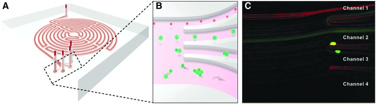
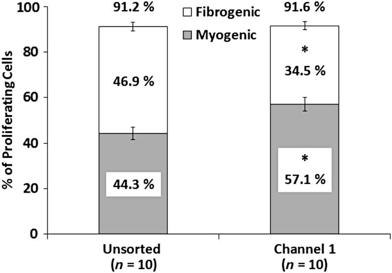
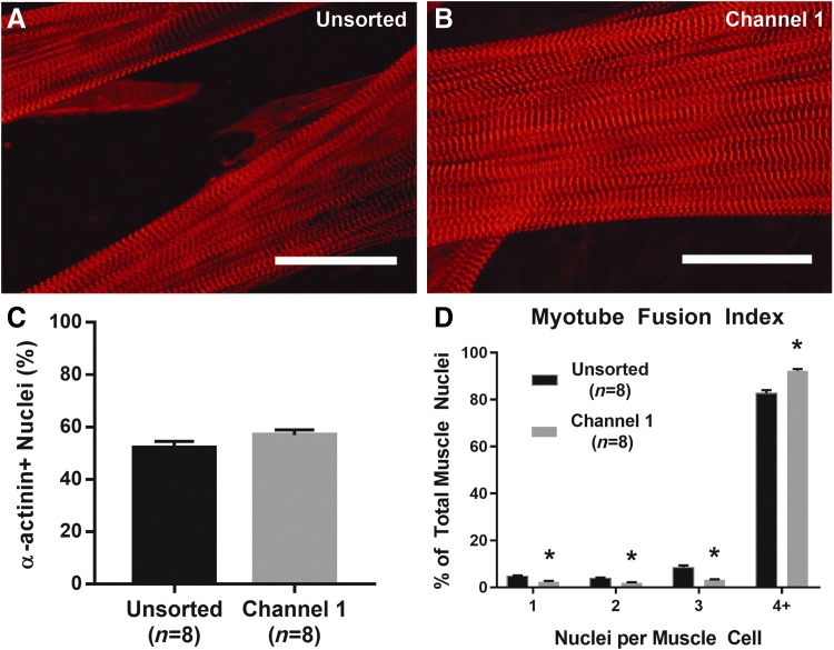
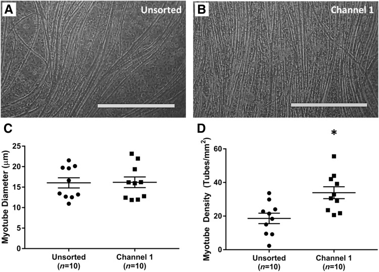
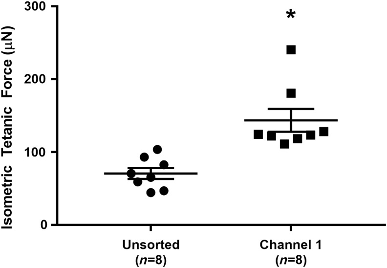
Skeletal muscle satellite cells have tremendous therapeutic potential in cell therapy or skeletal muscle tissue engineering. Obtaining a sufficiently pure satellite cell population, however, presents a significant challenge. We hypothesized that size differences between satellite cells and fibroblasts, two primary cell types obtained from skeletal muscle dissociation, would allow for label-free, inertial separation in a microfluidic device, termed a "Labyrinth," and that these purified satellite cells could be used to engineer skeletal muscle. Throughout tissue fabrication, Labyrinth-purified cells were compared with unsorted controls to assess the efficiency of this novel sorting process and to examine potential improvements in myogenic proliferation, differentiation, and tissue function. Immediately after dissociation and Labyrinth sorting, cells were immunostained to identify myogenic cells and fibroblast progenitors. Remaining cells were cultured for 14 days to form a confluent monolayer that was induced to delaminate and was captured as a 3D skeletal muscle construct. During monolayer development, myogenic proliferation (BrdU assay on Day 4), differentiation and myotube fusion index (α-actinin on Day 11), and myotube structural development (light microscopy on Day 14) were assessed. Isometric tetanic force production was measured in 3D constructs on Day 16. Immediately following sorting, unsorted cells exhibited a myogenic purity of 39.9% ± 3.99%, and this purity was enriched approximately two-fold to 75.5% ± 1.59% by microfluidic separation. The BrdU assay on Day 4 similarly showed significantly enhanced myogenic proliferation: in unsorted controls 47.0% ± 2.77% of proliferating cells were myogenic, in comparison to 61.7% ± 2.55% following purification. Myogenic differentiation and fusion, assessed by fusion index quantification, showed improvement from 82.7% ± 3.74% in control to 92.3% ± 2.04% in the purified cell population. Myotube density in unsorted controls, 18.6 ± 3.26 myotubes/mm2, was significantly enriched in the purified cell population to 33.9 ± 3.74 myotubes/mm2. Constructs fabricated from Labyrinth-purified cells also produced significantly greater tetanic forces (143.6 ± 16.9 μN) than unsorted controls (70.7 ± 8.03 μN). These results demonstrate the promise of microfluidic sorting in purifying isolated satellite cells. This unique technology could assist researchers in translating the regenerative potential of satellite cells to cell therapies and engineered tissues.

Keywords: microfluidics; optical imaging; skeletal muscle; tissue engineering; two-photon.


Conflict of interest statement

The authors have no competing financial interests to disclose.

Figures

<b>FIG. 1.</b>
FIG. 1.
Microfluidic Inertial Separation in the Labyrinth Device. A schematic representation of the Labyrinth is shown in (A). Cells in suspension enter the device at the top of the image and rapidly flow (1800 μL/min) along the circuitous path created by a series of curved channels. Dean forces proportional to cell size and channel curvature act on the cells transversely to the flow direction, separating distinct size classes of particles at the outlets as pictured in (B). Specifically, we intended to separate satellite cells, with a size range of 8–13 μm, from myofibroblasts, with a typical diameter of 10–22 μm. The Labyrinth was designed to focus the smaller satellite cells into Channel 1 (top), larger myofibroblasts into Channel 2 (second from top), and cell aggregates and debris into Channels 3 and 4 (bottom). (C) Visualization of a mixed population of Pax7+ satellite cells expressing red tdTomato fluorescence and Achilles tendon fibroblasts labeled with CellTracker Green fluorescent dye during sorting confirmed efficient separation of these two cell types based on their difference in size.
<b>FIG. 2.</b>
FIG. 2.
Myogenic and Fibrogenic Proliferation of Sorted Cells. Incorporation of BrdU on Day 4 of SMU fabrication was used to identify proliferating cells. No difference in overall proliferation was observed between unsorted controls (91.2% ± 1.2%) and cells sorted into Channel 1 (91.6% ± 1.0%, p = 0.556), suggesting the sorting process did not adversely affect cell growth. Immunostaining for MyoD and FSP1 indicated myogenic and fibrogenic cells, respectively. Proliferating myogenic cells were significantly enriched (p = 0.004) after sorting into Channel 1 (57.1% ± 3.0%) as compared with unsorted controls (44.3% ± 2.8%). In contrast, FSP1 staining indicated by proliferating fibrogenic cells in unsorted controls, 46.9% ± 1.9%, were significantly decreased to 34.5% ± 1.9% by microfluidic sorting (p < 0.001). * Indicates statistical difference from control.
<b>FIG. 3.</b>
FIG. 3.
Structural Maturation following Microfluidic Sorting. (A, B) Advanced sarcomeric structure within highly aligned myofibrils, evident from immunostaining for α-actinin, was observed on Day 11 in both unsorted controls and Channel 1 plates. Scale Bar = 50 μm. (C) No significant difference was recorded in the total nuclei associated with α-actinin-positive muscle cells (p = 0.142). (D) Quantification of myotube fusion index, the percentage of muscle cells with either 1, 2, 3, or 4+ nuclei, indicated greater fusion following microfluidic sorting. In particular, the percentage of fully fused myotubes with 4+ nuclei significantly increased from 82.7% ± 3.7% in unsorted controls to 92.3% ± 2.0% in Channel 1 (p < 0.001). * Indicates statistical difference from control.
<b>FIG. 4.</b>
FIG. 4.
Effects of Microfluidic Sorting on Myotube Growth. Shortly before delamination, light microscopy images were captured on Day 14 to assess the size and density of myotubes within the developing muscle monolayer. (A, B) Representative images of monolayers from unsorted cells and cell sorted into Channel 1. Scale Bar = 500 μm. (C) The average myotube diameter was indistinguishable between the two groups: 16.0 ± 1.3 μm in controls and 16.18 ± 1.4 μm in Channel 1 (p = 0.938). (D) Channel 1 cells, however, exhibited a significantly denser myotube network (p = 0.004), with 33.9 ± 3.7 tubes/mm2 in comparison to 18.6 ± 3.3 myotubes/mm2 in unsorted controls. * Indicates statistical difference from control.
<b>FIG. 5.</b>
FIG. 5.
SMU Functional Development with Microfluidic Sorting. Functional measurement of isometric tetanic force in 3D SMUs on Day 16 indicated a significant effect of sorting: unsorted control SMU force production of 71 ± 8 μN was significantly increased (p = 0.002) approximately two-fold to 144 ± 17 μN in Channel 1 SMUs. * Indicates statistical difference from control. SMU, skeletal muscle unit.

Similar articles

Cited by

References

    1. Järvinen T.A.H., Järvinen T.L.N., Kääriäinen M., Kalimo H., and Järvinen M. Muscle injuries: biology and treatment. Am J Sports Med 33, 745, 2005 - PubMed
    1. Turner N., and Badylak S. Regeneration of skeletal muscle. Cell Tissue Res 347, 759, 2012 - PubMed
    1. Huard J., Li Y., and Fu F.H. Muscle injuries and repair: current trends in research. J Bone Joint Surg 84, 822, 2002 - PubMed
    1. Shadrach J.L., and Wagers A.J. Stem cells for skeletal muscle repair. Philos Trans R Soc Lond B Biol Sci 366, 2297, 2011 - PMC - PubMed
    1. Tedesco F.S., Dellavalle A., Diaz-Manera J., Messina G., and Cossu G. Repairing skeletal muscle: regenerative potential of skeletal muscle stem cells. J Clin Invest 120, 11, 2010 - PMC - PubMed

Publication types

LinkOut - more resources