. 2017 Nov 6;214(11):3361-3379.
doi: 10.1084/jem.20161564. Epub 2017 Sep 25.

Spatially restricted JAG1-Notch signaling in human thymus provides suitable DC developmental niches

Affiliations

Spatially restricted JAG1-Notch signaling in human thymus provides suitable DC developmental niches

Enrique Martín-Gayo et al. J Exp Med. .

Abstract

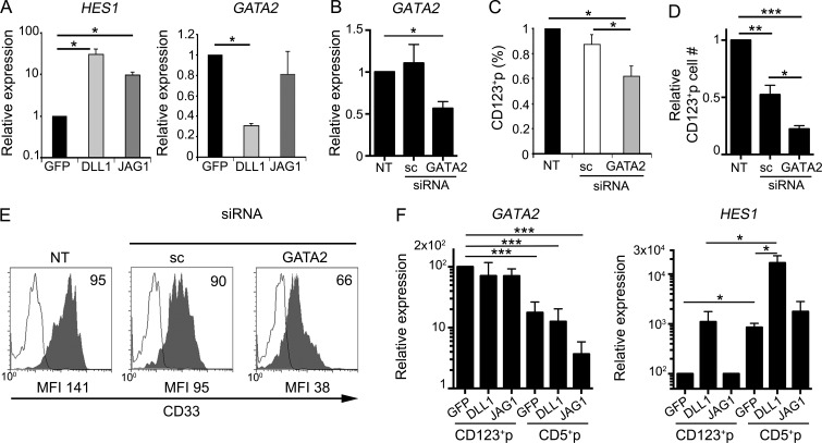
A key unsolved question regarding the developmental origin of conventional and plasmacytoid dendritic cells (cDCs and pDCs, respectively) resident in the steady-state thymus is whether early thymic progenitors (ETPs) could escape T cell fate constraints imposed normally by a Notch-inductive microenvironment and undergo DC development. By modeling DC generation in bulk and clonal cultures, we show here that Jagged1 (JAG1)-mediated Notch signaling allows human ETPs to undertake a myeloid transcriptional program, resulting in GATA2-dependent generation of CD34+ CD123+ progenitors with restricted pDC, cDC, and monocyte potential, whereas Delta-like1 signaling down-regulates GATA2 and impairs myeloid development. Progressive commitment to the DC lineage also occurs intrathymically, as myeloid-primed CD123+ monocyte/DC and common DC progenitors, equivalent to those previously identified in the bone marrow, are resident in the normal human thymus. The identification of a discrete JAG1+ thymic medullary niche enriched for DC-lineage cells expressing Notch receptors further validates the human thymus as a DC-poietic organ, which provides selective microenvironments permissive for DC development.


Figures

Figure 1.
Figure 1.
Human ETPs display monocyte/DC potential and generate in vitro pDCs and cDCs that resemble their intrathymic counterparts. (A) Flow cytometry phenotype of ETPs (CD34hi CD44hi CD33+ CD1a) isolated from the human postnatal thymus. Numbers indicate percentages of positive cells for the indicated markers (n = 20). (B) Flow cytometry of cells generated from human ETPs cultured on OP9 cell monolayers in the presence of 100 IU/ml rhFLT3L and 200 IU/ml rhIL-7 (OP9 assay) at the indicated days. Numbers correspond to percentages of cells that display either a pDC (BDCA2+ CD123hi CD13) or a cDC (BDCA2 CD123lo CD13hi) phenotype. Expression of CD1a, CD33, CD11c, and BDCA1 on cDCs and HLA-DR expression on cDCs and pDCs are also shown (n = 12). (C) Kinetics of pDC and cDC differentiation from ETPs cultured as in B. Data are shown as mean ± SEM of percentages (left) and absolute numbers (right) of pDCs and cDCs recovered at the indicated days of culture and normalized to 105 initial ETPs (n = 12). *, P < 0.05; **, P < 0.01. (D) Generation of monocytes from human ETPs cultured in the OP9 assay either as in B, or in the presence of M-CSF, for 7 d. Flow cytometry plots show the expression of CD115 and CD14 on electronically gated ETP-derived CD123lo CD13+ progenitor cells (shaded histograms). Background staining was determined with irrelevant isotype-matched Abs (empty histograms). Data are representative of one out of three experiments.
Figure 2.
Figure 2.
ETPs resident in the human thymus generate pDCs and cDCs in vivo. (A) Flow cytometry plots show expression of CD7 (left) and CD5 (right) either on primary pDCs (top) and cDCs (bottom) resident in the human thymus and in peripheral blood (PB) or on their DC counterparts generated in vitro from ETPs in the OP9 assay. Data are representative of one of three experiments. (B) Percentages of pDCs and cDCs among human CD45+ cells recovered from the spleen and BM of Rag 2−/− × γc−/− mice transplanted by intrahepatic injection with 2–5 × 10⁵ human ETPs. Percentages of total human CD45+ cells arising in the thymus are shown in the far-right graph. Mean values at 1 wk (n = 6) and 2 wk (n = 3) after transplantation are shown. (C) Flow cytometry analysis of human cells generated in mice transplanted with human ETPs at 1 wk after transplant. Numbers indicate percentages of CD45+ cells that display either a pDC (BDCA2+ CD123hiCD13) or a cDC (BDCA2 CD123lo CD13hi) phenotype in the spleen and BM or percentages of CD5+ CD13 T-lineage cells developing in the thymus.
Figure 3.
Figure 3.
Human ETPs differentiate in vitro into separate progenitors transcriptionally primed for either a lymphoid/T or a myeloid cell fate. (A and B) Phenotype of cells generated from human ETPs cultured in the OP9 assay for 3 d. (A) Flow cytometry plots show two exclusive cell subsets defined by reciprocal expression of CD5 and CD123 as CD5+ CD123 (CD5+p) and CD5lo CD123+ (CD123+p), which lack pDC (BDCA2) and cDC (CD11c) markers but express low CD13. (B) Flow cytometry plots of electronically gated CD5+p (gate I) and CD123+p (gate II) subsets in A show expression of CD117 and CD135 cytokine receptors together with CD34 and CD45RA, a phenotype of hematopoietic progenitors. Expression of CD115 on CD123+p identifies progenitors of monocytes and DCs (MDPs), while CD116+ CD115 CD123+p correspond to CDPs with the capacity to give rise to all DCs but not to monocytes (Breton et al., 2015b). Background was defined using isotype-matched irrelevant Abs (empty histograms). Representative results from one experiment are shown (n = 9). (C) Quantitative PCR expression analysis of the indicated genes in primary ETPs, pDCs, and cDCs purified from human postnatal thymus samples and in FACS-sorted CD5+p and CD123+p ETP-derived progenitors shown in A. Data were normalized to GAPDH expression. All results are shown relative to those of ETP as mean ± SEM (n = 3). *, P < 0.05.
Figure 4.
Figure 4.
Human ETPs generate pDCs and cDCs through myeloid-primed CD123+ CDPs. (A and B) Flow cytometry analysis of surface CD5 versus CD123 expression on FACS-sorted CD5+p (A) and CD123+p (B) progenitor subsets generated from human ETPs cultured for 3 d in the OP9 assay, as shown in Fig. 3 A. (C and D) Phenotype of cells generated from FACS-sorted CD5+p (C) and CD123+p (D) progenitors upon culture for 3 additional days in the OP9 assay. CD123+p progenitors generated pDCs (CD123+ CD13) and cDCs (CD123 CD13+), whereas CD5+p progenitors exclusively generated T-lineage cells (CD5+ CD7+ CD4+ CD8+). (E) Differentiation kinetics of pDCs (top), cDCs (middle), and T-lineage cells (bottom), derived from either CD123+p or CD5+p sorted progenitors upon culture as in A for 3 and 6 additional days, respectively. Mean percentages ± SEM are shown (n = 8). **, P < 0.01.
Figure 5.
Figure 5.
Characterization of CD123+ MDPs and CDPs resident in the human thymus. (A) Flow cytometry of CD44 versus CD34 expression in human thymocytes depleted of T-lineage cells, as described in Materials and methods, defines three progenitor populations: (I) CD44hi CD34hi ETPs, (II) CD34lo CD44hi myeloid-like intrathymic progenitors, and (III) CD34lo CD44lo lymphoid-like intrathymic progenitors. Shaded histograms show expression of the indicated markers on electronically gated subsets I, II, and III (bottom). Isotype-matched irrelevant Abs were used to define background staining (empty histograms; n = 3). According to their phenotype, cells in gate II resemble CDPs and also include some CD115+ MDPs. (B) Flow cytometry plots show the phenotype of myeloid progenitors (gate II) that were magnetically sorted on the basis of CD123 expression. Data from a representative experiment are shown (n = 4). (C) Kinetics of pDC and cDC differentiation from ETPs and primary CD123+ myeloid progenitors shown in B, isolated from the same thymus sample and cultured in the OP9 assay as in Fig. 1 B. Data are shown as mean percentages ± SEM (n = 4). *, P < 0.05.
Figure 6.
Figure 6.
The human thymus contains a JAG1+ medullary niche enriched for pDCs and cDCs expressing NOTCH4. (A–C) Immunohistochemistry and confocal microscopy analysis of JAG1 (red) and CD11c (green) expression in the human postnatal thymus. Topro labeling of cellular nuclei is shown in blue. Bars: 50 μm (A and B); 10 μm (C). (A) Thymus cortex (c) and medulla (m) are separated by the corticomedullary junction (dotted line). (B) Cell contacts between CD11c+ DCs and JAG1+ stromal cells confined to the thymus medulla (arrowheads). (C) Enlarged details of cell contacts between CD11c+ and JAG1+ cells in the thymus medulla marked by arrowheads. (D) Quantification of CD11c+ cells contacting JAG1+ cells in C. Data show mean percentages ± SEM per 63× field of three independent thymus samples (n = 30). (E) Quantitative PCR expression analysis of NOTCH (1–4) transcription in primary ETPs, pDCs, and cDCs purified from human postnatal thymus samples and in FACS-sorted CD5+p and CD123+p ETP-derived progenitors shown in Fig. 4 (A and B), respectively. Data were normalized to GAPDH expression. All results are shown relative to those of ETP as mean ± SEM (n = 3). *, P < 0.05. (F) Percentages of cells in E positive by flow cytometry for the expression of NOTCH1, 3, and 4. Data are shown as mean ± SEM of cell percentages (n = 3). *, P < 0.05; **, P < 0.01. (G) Flow cytometry analysis of cell surface NOTCH1, 3, and 4 expression on primary ETPs, CD5+p and CD123+p cells isolated from the human thymus (shaded histograms). Gray dotted histograms represent background staining obtained with irrelevant isotype-matched Abs. (H–J) Immunohistochemistry and confocal microscopy analysis of NOTCH4 protein expression (red) in combination with either CD123 or CD11c (green) in the human postnatal thymus. (H′–J′) Topro labeling of cellular nuclei is shown in blue. Bars, 50 μm. (H) NOTCH4 expression is confined to the human thymus medulla. Thymus cortex (c) and medulla (m) are separated by the corticomedullary junction (dotted line). (I and J) Enlarged details of the corticomedullary junction (CMJ) area of human thymus showing expression of NOTCH4 on CD123+ (I) and CD11c (J) DCs (arrowheads) confined to the medulla, close to the perivascular space (dotted lines).
Figure 7.
Figure 7.
JAG1- but not DLL1-mediated Notch signaling supports the generation of CD123+ thymic CDPs from human CD7, CD7hi, and total ETPs. (A and D) Percentages of either total DCs, including pDCs and cDCs (left), or CD5+ T-lineage cells (right), derived from total ETPs (A) or CD7 ETPs (D), cultured onto OP9–GFP, OP9–DLL1, or OP9–JAG1 stromas, in the presence of FLT3L and IL-7 (n = 3). (B, E, and H) Percentages of CD123+ myeloid progenitors, mostly including CDPs as shown in Fig. 3, arising from human total (B), CD7 (E), or CD7hi ETPs (H), cocultured for the indicated days with OP9–DLL1, OP9–JAG1, or OP9–GFP stromal cells as in A (n = 3). (C and G) Flow cytometry phenotype (left) and relative (middle) and absolute (right) numbers of pDCs and cDCs derived from 105 FACS-sorted CD7 (C) or CD7hi (G) ETPs in the OP9 assay, at the indicated days. Numbers in quadrants correspond to percentages of gated cells that display either a pDC (CD123hi CD13) or a cDC (CD123lo CD13hi) phenotype after 9 d of culture (n = 3 or 4). (F) Numbers of CD123+ myeloid progenitors generated from human CD7 ETPs in OP9–JAG1 cultures supplemented with either 100 nM of the gamma-secretase inhibitor (GSI) CompE or DMSO as a vehicle. Cell numbers in GSI cultures were normalized to those recovered in DMSO cultures (n = 3). Data in A–H are shown as mean ± SEM values. *, P < 0.05; ** P < 0.01; ***, P < 0.001.
Figure 8.
Figure 8.
Single CD7 ETPs generate both dendritic cells and T-lineage cells in vitro. (A) Phenotype of myeloid/DC- and T-lineage cells generated at the indicated days in bulk cultures from human CD7 ETPs (105 cells) seeded onto OP9–JAG1 or OP9–DLL1 cells in the presence of FL3TL and IL-7. Numbers in quadrants represent percentages of pDCs (CD123hi CD13), cDCs (CD13hiCD123lo), T-lineage (CD7+CD5+), and CD7+CD5 progenitors. (B) Representative confocal microscopy images of the clonal progeny obtained from culturing individual CD7 ETPs in OP9–JAG1 and OP9–DLL1 cultures at day 9 or 14, respectively. T-lineage cells were identified among CD45+ hematopoietic cells (blue) by exclusive expression of CD5 (red). Myeloid/DC-lineage cells were detected by coexpression of CD45 and CD13 and/or CD123 (yellow). OP9 cells expressing GFP are shown in green. Original magnification 20×. Scale bar is shown. (C) Clonal efficiency calculated on the basis of the number of positive wells is indicated. (D) Graphs represent the cellular output of all positive single cell cultures of CD7 ETPs. The number of wells per category is indicated at the top of each bar. My/DC, myeloid/DC-lineage (CD45+ CD13/CD123+); T, T-lineage (CD45+ CD13/CD123 CD5+); Undiff, undifferentiated cells (CD45+ CD13/CD123 CD5).
Figure 9.
Figure 9.
JAG1- but not DLL1-mediated Notch signaling supports a GATA2-dependent generation of CD123+ thymic CDPs from human ETPs. (A) Quantitative PCR analysis of HES1 (left) and GATA2 (right) gene expression in total ETPs cultured for 24 h with OP9–DLL1, OP9–JAG1, or OP9–GFP cells, in the presence of FLT3L and IL-7. Data were normalized to GAPDH expression. Results are shown as mean ± SEM expression values normalized to those of ETPs cultured onto OP9–GFP controls (n = 3). *, P < 0.05. (B) Relative expression of GATA2 in human ETPs that were either nucleofected with GATA2-specific or scramble (sc) siRNAs or nontransfected (NT) and then cultured onto OP9–GFP cells for 3 d in the presence of FLT3L and IL-7. Data were normalized to GAPDH expression and are shown as mean ± SEM values normalized to those of nontransfected (NT) ETPs (n = 3). *, P < 0.05. (C and D) Numbers of CD123+ CDPs generated as shown in B from human ETPs nucleofected with GATA2 or sc siRNAs. Data are represented as mean ± SEM percentages (C) or absolute numbers (D) of cells normalized to those of nontransfected ETPs (NT; n = 3). *, P < 0.05; **, P < 0.01; ***, P < 0.001. (E) Flow cytometry histograms show CD33 expression levels (shaded) of CD123+ CDPs in C and D. Background was determined using isotype-matched irrelevant Abs (empty histograms). Numbers indicate percentages of positive cells. Mean fluorescence intensity values are indicated at the bottom. (F) Quantitative PCR analysis of relative expression of GATA2 and HES1 transcripts in cells derived from FACS-sorted CD123+p or CD5+p subsets shown in Fig. 4 B that were cultured for 48 additional hours onto OP9–DLL1 or OP9–JAG1. Data are normalized to GAPDH expression and are shown as mean ± SEM values normalized to those of CD123+p cultured on OP9–GFP control stromal cells (n = 3). *, P < 0.05; ***, P < 0.001.
Figure 10.
Figure 10.
Both DLL1- and JAG1-mediated Notch signaling support the survival of intrathymic CD123+ CDPs and their development into pDCs and cDCs. (A) Flow cytometry analysis of CD123 versus CD13 expression on the cell progeny of FACS-sorted CD123+ CDPs derived from ETPs as shown in Fig. 4 B (CD123+p), which were cultured for 3 additional days onto OP9–GFP cells. The electronic gate defines the original CD123+ CD13lo/- phenotype of the CD123+p cultured population. (B) Relative cell numbers of the CD123+p cell progeny gated as in A, recovered at the indicated days upon coculture with OP9–DLL1, OP9–JAG1, or OP9–GFP stroma. Data are shown as mean ± SEM of cell numbers normalized to those of OP9–GFP cultures (n = 7). *, P < 0.05. (C) Proliferation kinetics analyzed by CFSE labeling and flow cytometry of FACS-sorted CD123+p shown in Fig. 4 B, and cultured as in B. Bars and error bars are MFI ± SEM,(n = 3). *, P < 0.05. (D) Flow cytometry analysis of apoptotic cells (shaded histograms) recovered at the indicated days from cultures in B. Numbers show percentages of Annexin V+ apoptotic cells. Empty histograms show background staining. Data correspond to a representative experiment (n = 3). (E) Absolute numbers of cells derived from primary CD123+ CDPs, isolated from the human thymus as shown in Fig. 5, which were cultured for the indicated days on OP9 stroma in the presence of either 100 nM of CompE (GSI) or DMSO as control. Bars and error bars are mean ± SEM (n = 3). *, P < 0.05. (F) Relative numbers of cDCs (left) and pDCs (right) generated from intrathymic CD123+ CDPs in cultures shown in E. Data are shown as mean ± SEM of cell numbers in GSI cultures normalized to those in DMSO cultures (n = 3). *, P < 0.05; **, P < 0.01. (G) Absolute numbers of cDCs (left) and pDCs (right) generated from primary thymic CD123+ CDPs, cultured on OP9–DLL1, OP9–JAG1, or OP9–GFP stromas for the indicated days. Data are shown as mean ± SEM cell numbers normalized to 105 input CDPs (n = 7). *, P < 0.05.

Similar articles

Cited by

References

    1. Ardavin C., Wu L., Li C.L., and Shortman K.. 1993. Thymic dendritic cells and T cells develop simultaneously in the thymus from a common precursor population. Nature. 362:761–763. 10.1038/362761a0 - DOI - PubMed
    1. Banchereau J., and Steinman R.M.. 1998. Dendritic cells and the control of immunity. Nature. 392:245–252. 10.1038/32588 - DOI - PubMed
    1. Bell J.J., and Bhandoola A.. 2008. The earliest thymic progenitors for T cells possess myeloid lineage potential. Nature. 452:764–767. 10.1038/nature06840 - DOI - PubMed
    1. Bendriss-Vermare N., Barthélémy C., Durand I., Bruand C., Dezutter-Dambuyant C., Moulian N., Berrih-Aknin S., Caux C., Trinchieri G., and Brière F.. 2001. Human thymus contains IFN-alpha-producing CD11c(-), myeloid CD11c(+), and mature interdigitating dendritic cells. J. Clin. Invest. 107:835–844. 10.1172/JCI11734 - DOI - PMC - PubMed
    1. Blom B., and Spits H.. 2006. Development of human lymphoid cells. Annu. Rev. Immunol. 24:287–320. 10.1146/annurev.immunol.24.021605.090612 - DOI - PubMed

MeSH terms