. 2017 Sep 25;7(1):12249.
doi: 10.1038/s41598-017-12527-9.

Human Cerebrospinal fluid promotes long-term neuronal viability and network function in human neocortical organotypic brain slice cultures

Affiliations

Human Cerebrospinal fluid promotes long-term neuronal viability and network function in human neocortical organotypic brain slice cultures

Niklas Schwarz et al. Sci Rep. .

Abstract

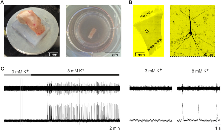
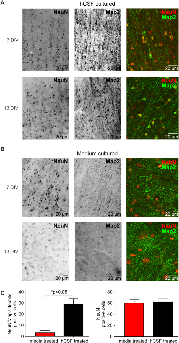
Pathophysiological investigation of CNS-related diseases, such as epilepsy or neurodegenerative disorders, largely relies on histological studies on human post mortem tissue, tissue obtained by biopsy or resective surgery and on studies using disease models including animal models, heterologous expression systems or cell culture based approaches. However, in general it remains elusive to what extent results obtained in model systems can be directly translated to the human brain, calling for strategies allowing validation or even primary investigation in live human CNS tissue. In the work reported here, we prepared human organotypic slice cultures from access tissue of resective epilepsy surgery. Employing different culture conditions, we systematically compared artificial culturing media versus human cerbrospinal fluid (hCSF) obtained from patients with normal pressure hydrocephalus (NPH). Presented data demonstrates sustained cortical neuronal survival including not only maintenance of typical cellular electrophysiological properties and activity, such as robust action potential generation and synaptic connectivity, but also preservation of tonic and phasic network activity up to several weeks in vitro. As clearly delineated by immunocytochemistry, single cell patch clamp and extracellular recordings, we find that in contrast to artificial culturing media, hCSF significantly enhances neuron viability and maintenance of network activity.


Conflict of interest statement

The authors declare that they have no competing interests.

Figures

Figure 1
Figure 1
Human organotypic slices were cultured in six well plates for up to 21 days. (A) Slices were cut from tissue blocks perpendicular to the cortical surface (250–300 µm thick) and cultured in six well plates either in traditional culture media or human cerebrospinal fluid (hCSF). (B) Typical example of a human organotypic slice and a reconstruction of a neuron filled with biocytin after whole cell patch clamp recording revealing a pyramidal morphology (C) Multi-Unit extracellular population recording of a human organotypic slice culture, note the induction of rhythmic network discharges after increasing the extracellular K+ concentration to 8 mM (4 DIV). Boxes are magnified at the right.
Figure 2
Figure 2
Electrophysiological recordings in human organotypic slice cultures. (A) In human organotypic slice cultures three main activity patterns were observed: Rhythmic network activity (A, left panel, 13 DIV), tonic activity (A right panel, 14 DIV) or no activity at all (D upper panels, 7 DIV and 14–21 DIV). (B) Overall, the number of slices being able to produce neuronal activity was much higher for slices cultured in hCSF (left pie chart) compared to slices cultured in traditional media (right pie chart). (C) Slices were recorded after different days in vitro (3–21 DIV). Slices cultured in hCSF (black circles) very frequently exhibited activity in comparison to slices cultured in traditional media (red triangles) (C and D). During the first days in vitro slices cultured in traditional media were still able to produce rhythmic and tonic activity, while in slices cultured for more than 7–10 days no rhythmic activity and just very rarely tonic activity could be recorded. In contrast, the majority of slices cultured in hCSF for 3–21 days showed either rhythmic or tonic activity (B,C and D).
Figure 3
Figure 3
Rhythmic network activity of human slice cultures. The slices that produced rhythmic activity showed either long lasting network discharges (A and E) or oscillations with shorter bursts occurring with higher frequencies in the presence of 8 mM K+ (D). Shown are examples of the Multi-Unit Activity (MUA) recorded with an extracellular electrode and a simultaneous intracellular recording of a cortical neuron in high potassium alone (A) and in high potassium and the presence of Muscarine 5 µM (B). The red insets in A and B show expansions of the traces above. (A) Note that the action potential firing is riding on a barrage of excitatory postsynaptic potential (EPSP). The neuron in (B) was slightly hyperpolarized with a holding current of – 50 pA to emphasize the synaptic inputs. (C) Plot of the frequency of the oscillation and duration of all recorded network events in high potassium. (D) Plot of the frequency of network discharges against the DIV (left) and duration (right). (E) In a subset of the experiments (n = 3) the application of CNQX abolished the network discharges.
Figure 4
Figure 4
Representative examples of intracellular whole cell patch clamp recordings and biocytin filled morphology of human cortical neurons cultured in hCSF. In slices cultured in hCSF non-pyramidal (A, 9 DIV) and pyramidal neurons (B, 2 DIV) could be filled with biocytin and the response to hyperpolarizing and depolarizing current injections could be recorded. (C) The majority of cells recorded in current clamp received spontaneous inhibitory and excitatory synaptic inputs. Red arrow marks an excitatory postsynaptic potential (EPSP) and blue arrow an inhibitory IPSP in a cell recorded after 13 DIV. (D) All cells recorded in hCSF-cultured slices showed sustained action potential generation due to positive current injections (+50 pA to +200 pA) and showed typical firing behavior as reported before in human non – pyramidal (9 DIV, D upper trace) or pyramidal neurons (2 DIV, D lower trace).
Figure 5
Figure 5
Effect of the culture medium on the excitability of human cortical neurons. The cortical cells recorded in slices cultured in hCSF (A) and after the first days in media (B, top) showed typical repetitive firing behavior to suprathreshold current injections (n = 10, 2–14 DIV for hCSF, n = 2, 3 DIV for media), while cells recorded from slices cultured in media for more than 7 days showed only a single action potential on suprathreshold current injections (B, bottom, n = 2).
Figure 6
Figure 6
Morphology of human cortical slices in culture. (A) Example of a human organotypic slice after 18 DIV, note the dense labeling of NeuN positive cells in superficial layers and deep layers of the cortex. (A1) Inset of the superficial layers which are enlarged in (B) showing intact morphology of Layer 2/3 NeuN positive neurons with typical morphology of pyramidal cells revealed by Map2 staining and overlay. (C) Representative example of the size of a typical slice culture after 12 DIV (5 mm × 3 mm) with white matter and layers I–VI. (D) GFAP positive labeled astrocytes with Map2 positiv pyramidal cells cultured in hCSF after 28 DIV.
Figure 7
Figure 7
Morphology of human cortical neurons of slices cultured in hCSF vs. traditional media. Typical example of a NeuN and Map2 staining of human organotypic slices cultured in hCSF after 7 and 13 DIV (A) or traditional media after 7 and 13 DIV (B). Confocal images of the same slice showing co-labeling of NeuN and Map2 and intact morphology of the cells in slices cultured in hCSF (A) and loss of dendritic morphology in slices cultured in traditional media (B). Scale bars in all panels represent 20 µm. (C) Quantification of NeuN/Map2 double positive neurons in slices treated in traditional media compared to hCSF.

References

    1. Kriks S, et al. Dopamine neurons derived from human ES cells efficiently engraft in animal models of Parkinson’s disease. Nature. 2011;480:547–551. - PMC - PubMed
    1. Zhang Y, et al. Rapid single-step induction of functional neurons from human pluripotent stem cells. Neuron. 2013;78:785–798. doi: 10.1016/j.neuron.2013.05.029. - DOI - PMC - PubMed
    1. Lancaster MA, et al. Cerebral organoids model human brain development and microcephaly. Nature. 2013;501:373−+. doi: 10.1038/nature12517. - DOI - PMC - PubMed
    1. D’Esposito M, Postle BR, Rypma B. Prefrontal cortical contributions to working memory: evidence from event-related fMRI studies. Experimental Brain Research. 2000;133:3–11. doi: 10.1007/s002210000395. - DOI - PubMed
    1. Clark RE, Squire LR. Classical conditioning and brain systems: the role of awareness. Science. 1998;280:77–81. doi: 10.1126/science.280.5360.77. - DOI - PubMed

Publication types