Ventricular myocardium development and the role of connexins in the human fetal heart
- PMID: 28947768
- PMCID: PMC5612926
- DOI: 10.1038/s41598-017-11129-9
Ventricular myocardium development and the role of connexins in the human fetal heart
Abstract
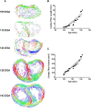
The developmental timeline of the human heart remains elusive. The heart takes on its characteristic four chambered appearance by ~56 days gestational age (DGA). However, owing to the complexities (both technical and logistical) of exploring development in utero, we understand little of how the ventricular walls develop. To address this, we employed diffusion tensor magnetic resonance imaging to explore the architecture and tissue organization of the developing heart aged 95-143 DGA. We show that fractional anisotropy increases (from ~0.1 to ~0.5), diffusion coefficients decrease (from ~1 × 10⁻³mm2/sec to ~0.4 × 10⁻³mm2/sec), and fiber paths, extracted by tractography, increase linearly with gestation, indicative of the increasing organization of the ventricular myocytes. By 143 DGA, the developing heart has the classical helical organization observed in mature mammalian tissue. This was accompanied by an increase in connexin 43 and connexin 40 expression levels, suggesting their role in the development of the ventricular conduction system and that electrical propagation across the heart is facilitated in later gestation. Our findings highlight a key developmental window for the structural organization of the fetal heart.
Conflict of interest statement
The authors declare that they have no competing interests.
Figures


Similar articles
-
Development of Helical Myofiber Tracts in the Human Fetal Heart: Analysis of Myocardial Fiber Formation in the Left Ventricle From the Late Human Embryonic Period Using Diffusion Tensor Magnetic Resonance Imaging.J Am Heart Assoc. 2020 Oct 20;9(19):e016422. doi: 10.1161/JAHA.120.016422. Epub 2020 Sep 30. J Am Heart Assoc. 2020. PMID: 32993423 Free PMC article.
-
Comparison of connexin 43, 40 and 45 expression patterns in the developing human and mouse hearts.Cell Commun Adhes. 2001;8(4-6):339-43. doi: 10.3109/15419060109080750. Cell Commun Adhes. 2001. PMID: 12064615
-
Comparison of connexin expression patterns in the developing mouse heart and human foetal heart.Mol Cell Biochem. 2003 Jan;242(1-2):121-7. Mol Cell Biochem. 2003. PMID: 12619874
-
Alterations in cardiac connexin expression in cardiomyopathies.Adv Cardiol. 2006;42:228-242. doi: 10.1159/000092572. Adv Cardiol. 2006. PMID: 16646594 Review.
-
Cardiac connexins: genes to nexus.Adv Cardiol. 2006;42:1-17. doi: 10.1159/000092550. Adv Cardiol. 2006. PMID: 16646581 Review.
Cited by
-
The within-subject application of diffusion tensor MRI and CLARITY reveals brain structural changes in Nrxn2 deletion mice.Mol Autism. 2019 Feb 28;10:8. doi: 10.1186/s13229-019-0261-9. eCollection 2019. Mol Autism. 2019. PMID: 30858964 Free PMC article.
-
Insoluble Aβ overexpression in an App knock-in mouse model alters microstructure and gamma oscillations in the prefrontal cortex, affecting anxiety-related behaviours.Dis Model Mech. 2019 Sep 24;12(9):dmm040550. doi: 10.1242/dmm.040550. Dis Model Mech. 2019. PMID: 31439589 Free PMC article.
-
Three-dimensional alignment of microvasculature and cardiomyocytes in the developing ventricle.Sci Rep. 2020 Sep 11;10(1):14955. doi: 10.1038/s41598-020-71816-y. Sci Rep. 2020. PMID: 32917915 Free PMC article.
-
Urolithin B, a Gut Microbiota Metabolite, Reduced Susceptibility to Myocardial Arrhythmic Predisposition after Hypoxia.Dis Markers. 2022 Feb 8;2022:6517266. doi: 10.1155/2022/6517266. eCollection 2022. Dis Markers. 2022. PMID: 35178131 Free PMC article.
-
Deficient Myocardial Organization and Pathological Fibrosis in Fetal Aortic Stenosis-Association of Prenatal Ultrasound with Postmortem Histology.J Cardiovasc Dev Dis. 2021 Sep 28;8(10):121. doi: 10.3390/jcdd8100121. J Cardiovasc Dev Dis. 2021. PMID: 34677190 Free PMC article.
References
-
- LeGrice IJ, et al. Laminar structure of the heart: ventricular myocyte arrangement and connective tissue architecture in the dog. Am. J. Physiol. Heart Circ. Physiol. 1995;269:H571–82. - PubMed
Publication types
MeSH terms
Substances
Grants and funding
LinkOut - more resources
Full Text Sources
Other Literature Sources
Molecular Biology Databases

