. 2017 Sep 25;7(1):12276.
doi: 10.1038/s41598-017-12519-9.

Determinants of the assembly and function of antibody variable domains

Affiliations

Determinants of the assembly and function of antibody variable domains

Eva Maria Herold et al. Sci Rep. .

Abstract

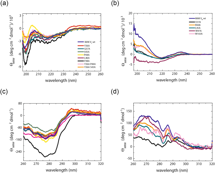
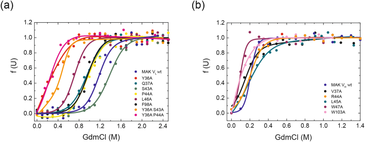
The antibody Fv module which binds antigen consists of the variable domains VL and VH. These exhibit a conserved ß-sheet structure and comprise highly variable loops (CDRs). Little is known about the contributions of the framework residues and CDRs to their association. We exchanged conserved interface residues as well as CDR loops and tested the effects on two Fvs interacting with moderate affinities (KDs of ~2.5 µM and ~6 µM). While for the rather instable domains, almost all mutations had a negative effect, the more stable domains tolerated a number of mutations of conserved interface residues. Of particular importance for Fv association are VLP44 and VHL45. In general, the exchange of conserved residues in the VL/VH interface did not have uniform effects on domain stability. Furthermore, the effects on association and antigen binding do not strictly correlate. In addition to the interface, the CDRs modulate the variable domain framework to a significant extent as shown by swap experiments. Our study reveals a complex interplay of domain stability, association and antigen binding including an unexpected strong mutual influence of the domain framework and the CDRs on stability/association on the one side and antigen binding on the other side.


Conflict of interest statement

The authors declare that they have no competing interests.

Figures

Figure 1
Figure 1
Conserved residues within the interface between VL and VH. In (a) the positions of conserved amino acids in the Fv fragment of MAK33 are shown. The VL domain is depicted in light green with the three CDRs highlighted in dark green. VH is shown in light blue and the CDRs are in dark blue. Conserved residues within the interface are illustrated as spheres and color-coded. On the right, the region within the rectangle is enlarged. For a better orientation CDR-H2 is indicated. The labelled residues were selected for an alanine-exchange mutational analysis. In (b) and (c) the top views of the interacting residues of the VL (left) and VH (right) domain of MAK33 (b) and 1HEZ (c) are shown. Structures are modified from PDB ID 1FH5 and 1HEZ. The grey dotted line indicates CDR-H3 which is not resolved in PDB ID 1FH5.
Figure 2
Figure 2
Secondary and tertiary structure of MAK33 VL and VH alanine exchange mutants. In (a) and (b), FUV-CD spectra of VL mutants (a) and VH mutants (b) are shown. Color code for VL: Y36A is red, Q37A dark cyan, S43A green, P44A yellow, L45A purple, F98A black, Y36A/S43A orange, Y36A/P44A pink and VL wild type is royal blue. Color code for VH: V37A is black, R44A orange, L45A dark cyan, W47A purple, W103A pink and VH wild type marine blue. In (c) and (d) the NUV-CD spectra of the VL (c) and VH (d) point mutants are shown. Color code for (c) and (d) is analogue to (a) and (b), respectively. For the spectra 16 accumulations each were recorded and buffer-corrected (PBS). All measurements were performed at a protein concentration of 20 µM (FUV-CD) and 50 µM (NUV-CD) in 0.5-mm (FUV) or 5-mm (NUV) quartz cuvette at 20 °C.
Figure 3
Figure 3
Secondary and tertiary structure of 1HEZ VL and VH alanine exchange mutants. In (a) and (b), FUV-CD spectra of VL mutants (a) and VH mutants (b) are shown. Color code for VL: Y36A is red, Q37A dark cyan, P44A yellow, L45A purple, F98A black, and VL wild type is royal blue. Color code for VH: V37A is black, G44A orange, L45A dark cyan, W47A purple, W103A pink and VH wild type marine blue. In (c) and (d) the NUV-CD spectra of the VL (c) and VH (d) point mutants are shown. Color code for (c) and (d) is analogue to (a) and (b), respectively. For the spectra 16 accumulations each were recorded and buffer-corrected (PBS). All measurements were performed at a protein concentration of 20 µM (FUV-CD) and 50 µM (NUV-CD) in 0.5-mm (FUV) or 5-mm (NUV) quartz cuvette at 20 °C.
Figure 4
Figure 4
Influence of conserved residues on MAK33 protein stability. The stability of the VH and VL alanine point mutants towards GdmCl-induced denaturation (a) and (b). (a) VL mutants: Y36A is red, Q37A dark cyan, S43A green, P44A yellow, L46A purple, F98A black, Y36A/S43A orange, Y36A/P44A pink and the wild type marine blue. (b) VH mutants: V37A is black, R44A orange, L45A dark cyan, W47A purple, W103A pink and the wild type royal blue.
Figure 5
Figure 5
Influence of conserved residues on 1HEZ protein stability. The stability of the VH and VL alanine point mutants towards GdmCl-induced denaturation (a) and (b). (a) VL mutants: Y36A is red, Q37A dark cyan, P44A yellow, L46A purple, F98A black and the wild type marine blue. (b) VH mutants: V37A is black, G44A orange, L45A dark cyan, W47A purple, W103A pink and the wild type royal blue.
Figure 6
Figure 6
Conserved interface residues with importance for VH/VL association. The top-views for MAK33 (left) and 1HEZ (right) VL and VH are shown and the residues with strong influence on association highlighted in red, those with mild influence highlighted in orange. For MAK33 the numbering of the strands (a, b, c, c′, c″, d, e, f, g) is also shown. Structures are modified from PDB ID 1FH5 and 1HEZ. The grey dotted line indicates CDR-H3 which is not resolved in PDB ID 1FH5.
Figure 7
Figure 7
Influence of conserved residues on antigen binding. To determine the functionality of the mutants an ELISA with Fv fragments was performed. In (a) the ELISA setup is shown. The biotinylated antigen (creatine kinase) was immobilized on streptavidin coated 96 well plates. Subsequently, 1:1 mixtures of the FLAG-tagged wild type domain and a corresponding mutant were added. Binding was detected via a peroxidase-conjugated anti –FLAG antibody. The peroxidase mediated conversion of the substrate ABTS was measured colorimetrical at 405 nm. For VL (a) and VH (b), titrations of Fvs containing either the wild type, a representative of an inactive, a low active or a mutant with similar activity as the wild type is shown. The absorption at 405 nm at the signal maximum was corrected for the signal of the variable labelled domains alone. Experiments were performed at 20 °C.
Figure 8
Figure 8
CDR-grafted mutants. (a) Schematic representation of CDR-grafted mutants. The framework for VL grafting mutants (left) was 1DH5, which is the most stable human consensus sequence VL domain (from HuCAL). The framework for VH grafting mutants (right) was 1DHU, which is the most stable human consensus sequence for VH domains. In (b and c), FUV-CD spectra of VL (left) and VH (right) CDR-grafted mutants are shown. The color code for VL is: MAK_1DH5 in red, 1DH5_MAK in blue; wild type 1DH5 is shown in green MAK33 in black. The color code for VH is: MAK_1DHU in red, 1DHU_MAK in blue, wild type 1DHU in green, MAK33 in black and MAK33 CDR3 1DHU in brown. In (d and e) NUV-CD spectra of the VL (left) and VH (right) point mutants are shown.
Figure 9
Figure 9
The influence of the CDRs on the stability of the variable domains. To assess the stability of CDR-grafted mutants, GdmCl-induced unfolding experiments were performed. Data for denaturant-induced transitions for VL (a) und VH (b) variants are shown. Data were evaluated according to a two-state unfolding model to obtain midpoints and cooperativity parameters of the transitions. Measurements were performed at 20 °C at a protein concentration of 1 μM.
Figure 10
Figure 10
Interface hydration near mutation as determined by MD simulations. The snapshots illustrate the water distribution within 7 Å of the mutation site. Protein chains are shown as cartoon (VL: green, VH: blue). Atoms within 7 Å of the mutation site are indicated as van der Waals spheres using atom color code for water molecules (and grey for protein atoms). For comparison the same regions are also shown for the wild type case (left panels). In case of the VH W103 mutation the average shift in backbone structure is illustrated (green cartoon) and compared to the wild type case (light blue).

References

    1. Chothia C, Novotny J, Bruccoleri R, Karplus M. Domain association in immunoglobulin molecules. The packing of variable domains. Journal of molecular biology. 1985;186:651–663. doi: 10.1016/0022-2836(85)90137-8. - DOI - PubMed
    1. Morea V, Tramontano A, Rustici M, Chothia C, Lesk AM. Conformations of the third hypervariable region in the VH domain of immunoglobulins. Journal of molecular biology. 1998;275:269–294. doi: 10.1006/jmbi.1997.1442. - DOI - PubMed
    1. Vargas-Madrazo E, Paz-Garcia E. An improved model of association for VH-VL immunoglobulin domains: asymmetries between VH and VL in the packing of some interface residues. Journal of molecular recognition: JMR. 2003;16:113–120. doi: 10.1002/jmr.613. - DOI - PubMed
    1. Bork P, Holm L, Sander C. The immunoglobulin fold. Structural classification, sequence patterns and common core. Journal of molecular biology. 1994;242:309–320. - PubMed
    1. Abhinandan KR, Martin AC. Analysis and prediction of VH/VL packing in antibodies. Protein engineering, design & selection: PEDS. 2010;23:689–697. doi: 10.1093/protein/gzq043. - DOI - PubMed

Publication types

MeSH terms

Substances

LinkOut - more resources