. 2017 Jul 25;8(36):60378-60389.
doi: 10.18632/oncotarget.19534. eCollection 2017 Sep 1.

NOTCH1 activates the Wnt/β-catenin signaling pathway in colon cancer

Affiliations

NOTCH1 activates the Wnt/β-catenin signaling pathway in colon cancer

Hideyuki Ishiguro et al. Oncotarget. .

Abstract

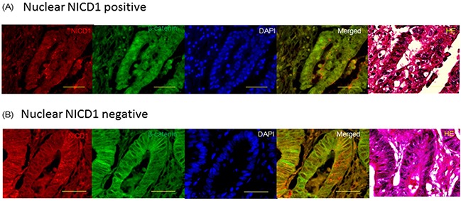
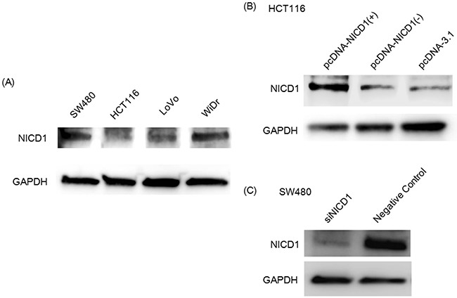
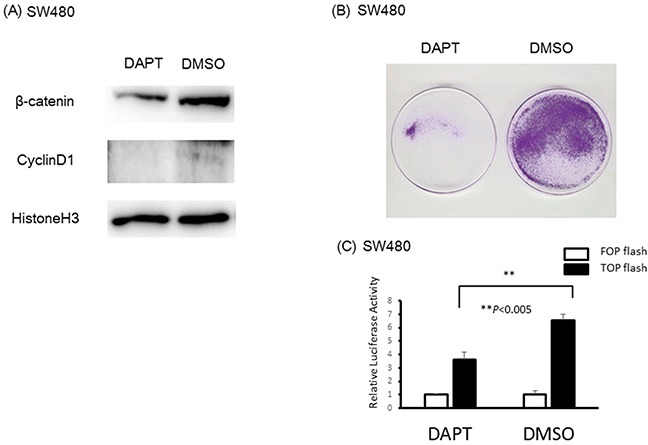
Purpose and methods: The translocation of β-catenin/CTNNB1 to the nucleus activates Wnt signaling and cell proliferation; however, the precise mechanism underlying this phenomenon remains unknown. Previous reports have provided evidence that NOTCH1 is involved in the Wnt signaling pathway. Therefore, we sought to determine the mechanism by which NOTCH1 influences the Wnt/β-catenin pathway. We constructed a vector expressing the NOTCH1 intracellular domain (NICD1) and transfected the vector into HCT116 which has low expression of NICD1. Furthermore, inhibition of NOTCH signal pathway in SW480 which has abundant NICD1 expression, was performed by transfection of siNICD1 or DAPT, gamma secretase inhibitor, treatment. In addition, we evaluated NICD1 and β-catenin localization in colon cancer cell lines and in 189 colon cancer tissue samples and analyzed the correlation between the nuclear localization of NICD1 and the clinicopathological features of colon cancer patients.

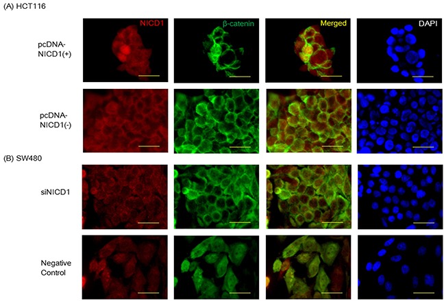
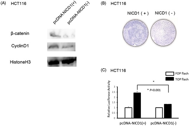
Results: Immunohistochemical assays demonstrated that NICD1 and β-catenin exhibited a similar localization pattern in colon cancer tissues. In addition, we found that NICD1 induced the translocation of β-catenin to the nucleus and that NICD1 and β-catenin co-localized in the nucleus. Overexpression of NICD1 increased luciferase activity of Wnt signal pathway. On the other hand, reduction of NICD1 reduced luciferase activity of Wnt signaling pathway. In the 189 analyzed colon cancer cases, multivariate COX regression analysis demonstrated the independent prognostic impact of nuclear localization of NICD1(p=0.0376).

Conclusion: NOTCH1 plays a key role in the Wnt pathway and activation of NOTCH1 is associated with the translocation of β-catenin to the nucleus.

Keywords: NICD1; NOTCH1; colon cancer; prognosis; β-catenin.


Conflict of interest statement

CONFLICTS OF INTEREST The authors have no proprietary or commercial interest in any product or concept discussed in this manuscript.

Figures

Figure 1
Figure 1. Confocal imaging of immunohistochemistry analysis of β-catenin and NICD1 in colon cancer tissues
Representative image of β-catenin and NICD1 localization in colon cancer tissue samples. DAPI, Merged image of NICD1 and β-catenin and HE stain. The antibody used (ab8925, from Abcam) actually recognizes the active form of Notch1 receptor, exposed after cleavage by γ-secretase. Yellow bar indicates 50um. (A) Nuclear NICD1 positive pattern: NICD1 and β-catenin were expressed in the nucleus of colon cancer cell. Merged image indicated that NICD1 and β-catenin were co-expressed in the same colon cancer cell. (B) Nuclear NICD1 negative pattern: NOTCH1 and β-catenin were expressed in the cytoplasm of colon cancer cell. Merged image indicated that NOTCH1 and β-catenin were co expressed in the same colon cancer cell.
Figure 2
Figure 2
(A) Wetern blot analysis of NICD1 expression in 4 colon cancer cell lines. The NICD1 band is approximately 120 kD. GAPDH was used as internal control. (B) Western blot analysis of HCT116 transfected with the pcDNA3.1-NICD1(+), pcDNA3.1-NICD1(-)(Mock) or pcDNA3.1 vector. (C) Western blotting analysis of NICD1 expression in siNICD1-transfected cells. NICD1 expression levels were suppressed in cells transfected with siNICD1 compared with Negative Control.
Figure 3
Figure 3
(A) Confocal imaging of NICD1 and β-catenin immunohistochemistry staining in HCT116 cells transfected with pcDNA3. 1NICD1(+) or pcDNA3.1-NICD1(-). In HCT116 cells transfected pcDNA3.1NICD1(+), β-catenin was robustly expressed. (B) Confocal imaging of NICD1 and β-catenin immunohistochemistry staining in SW480 cells transfected with siNICD1 or Negative Control. In SW480 cells transfected siNICD1, β-catenin expression in the nucleus was reduced. Yellow bar indicates 30um.
Figure 4
Figure 4. Induction of NICD1 into HCT cells with lower endogenous NICD1
(A) Western blotting analysis of NICD1, β-catenin and Cyclin D1 expression in pcDNA3.1-NICD1(+) or pcDNA3.1-NICD1(-)(Mock) -transfected cells. β-catenin and Cyclin D1 expression levels in the nucleus were increased in cells transfected with pcDNA3.1-NICD1(+). (B) Colony formation assay using HCT116 cells transfected with pcDNA3.1-NICD1(+) or pcDNA3.1-NICD1(-) (control). Colony formation in HCT116 cells transfected with pcDNA3.1-NICD1 was enhanced compared with HCT116 cells transfected with the control vector. (C) Relative β-catenin / TCF luciferase activity of HCT116 cells transfected with pcDNA3.1-NICD1(+) or pcDNA3.1-NICD1(-) (control).
Figure 5
Figure 5. Reduction of NICD1 into SW480 cells with abundant endogenous NICD1
(A) β-catenin and Cyclin D1 expression in the nucleus of SW480 cells treated with DAPT or DMSO(control). β-catenin and Cyclin D1 expression levels in the nucleus of SW480 cells were suppressed in cells transfected with DAPT. (B) Colony formation assay using SW480 cells treated with DAPT or DMSO (control). Colony formation in SW480 cells treated with DAPT was suppressed compared with SW480 cells treated with DMSO (control). (C) Relative β-catenin / TCF luciferase activity of SW480 cells treated with DAPT or DMSO (control).

References

    1. Nusse R, Varmus HE. Wnt genes. Cell. 1992;69:1073–1087. - PubMed
    1. van den Heuvel M, Nusse R, Johnston P, Lawrence PA. Distribution of the wingless gene product in Drosophila embryos: a protein involved in cell-cell communication. Cell. 1989;59:739–749. - PubMed
    1. Wieschaus E, Riggleman R. Autonomous requirements for the segment polarity gene armadillo during Drosophila embryogenesis. Cell. 1987;49:177–184. - PubMed
    1. Funayama N, Fagotto F, McCrea P, Gumbiner BM. Embryonic axis induction by the armadillo repeat domain of beta-catenin: evidence for intracellular signaling. J Cell Biol. 1995;128:959–968. - PMC - PubMed
    1. Nakamura Y. Cleaning up on beta-catenin. Nat Med. 1997;3:499–500. - PubMed

LinkOut - more resources