

Deceptive morphologic and epigenetic heterogeneity in diffuse intrinsic pontine glioma
- PMID: 28947983
- PMCID: PMC5601151
- DOI: 10.18632/oncotarget.19726
Deceptive morphologic and epigenetic heterogeneity in diffuse intrinsic pontine glioma
Abstract
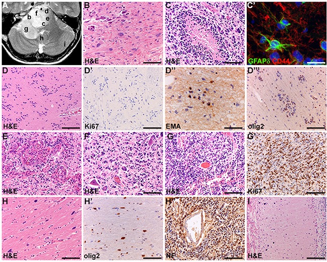
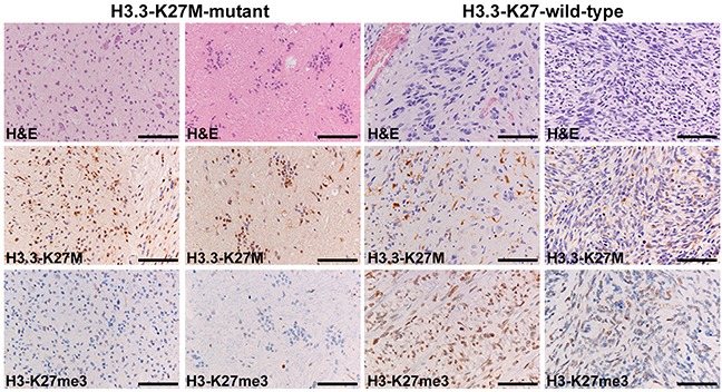
Historically, the diagnosis of diffuse intrinsic pontine glioma (DIPG) was based on typical imaging findings and clinical characteristics instead of pathology. However, the discovery of mutations in histone H3 variants, and the availability of tumor material for molecular analysis, has led to a paradigm shift in DIPG research and clinical practice. Using data from whole-brain autopsies in a series of nine DIPG patients with known histone mutational status, we here aim to review histopathological characteristics with special focus on intratumoral heterogeneity (ITH) and histone 3 K27 trimethylation (H3 K27me3). All DIPGs showed marked histologic ITH, with 56% even showing focal areas resembling a WHO grade I phenotype. As expected, H3 K27me3 immunoreactivity was lost in the tumors that were H3 K27M-mutated (seven patients; 67% H3.3, 11% H3.1). Strikingly, the H3K27 wildtype tumors (two patients; 22%) also contained H3 K27me3-immunonegative areas. Our study underscores the importance of the choice of the biopsy site, as ITH in DIPGs could theoretically lead to erroneous histological diagnoses with small biopsies. New in this respect is our finding that a substantial number of otherwise typical DIPGs has areas resembling WHO grade I tumors (esp. pilocytic astrocytoma, subependymoma). Furthermore, our study shows that negative H3 K27me3 immunohistochemistry in a DIPG does not imply a H3 K27-mutant tumor.
Keywords: H3 K27M; diffuse intrinsic pontine glioma; histone 3; intratumoral heterogeneity; trimethylation.
Conflict of interest statement
CONFLICTS OF INTEREST None.
Figures
References
-
- Epstein F. A staging system for brain stem gliomas. Cancer. 1985;56:1804–6. - PubMed
-
- Louis DN, Ohgaki H, Wiestler OD, Cavenee WK, Ellison DW, Figarella-Branger D, Perry A, Reifenberger G, Von Deimling A. WHO Classification of Tumours of the Central Nervous System. IARC Press; Lyon: 2016. - PubMed
-
- Caretti V, Jansen MH, van Vuurden DG, Lagerweij T, Bugiani M, Horsman I, Wessels H, van der Valk P, Cloos J, Noske DP, Vandertop WP, Wesseling P, Wurdinger T, et al. Implementation of a multi-institutional diffuse intrinsic pontine glioma autopsy protocol and characterization of a primary cell culture. Neuropathol Appl Neurobiol. 2013;39:426–36. - PubMed
LinkOut - more resources
Full Text Sources
Other Literature Sources
Research Materials
