. 2017 Sep 25;4(5):ENEURO.0248-17.2017.
doi: 10.1523/ENEURO.0248-17.2017. eCollection 2017 Sep-Oct.

NCS-Rapgef2, the Protein Product of the Neuronal Rapgef2 Gene, Is a Specific Activator of D1 Dopamine Receptor-Dependent ERK Phosphorylation in Mouse Brain

Affiliations

NCS-Rapgef2, the Protein Product of the Neuronal Rapgef2 Gene, Is a Specific Activator of D1 Dopamine Receptor-Dependent ERK Phosphorylation in Mouse Brain

Sunny Zhihong Jiang et al. eNeuro. .

Erratum in

Abstract

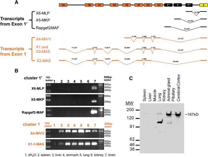
The neuritogenic cAMP sensor (NCS), encoded by the Rapgef2 gene, links cAMP elevation to activation of extracellular signal-regulated kinase (ERK) in neurons and neuroendocrine cells. Transducing human embryonic kidney (HEK)293 cells, which do not express Rapgef2 protein or respond to cAMP with ERK phosphorylation, with a vector encoding a Rapgef2 cDNA reconstituted cAMP-dependent ERK activation. Mutation of a single residue in the cyclic nucleotide-binding domain (CNBD) conserved across cAMP-binding proteins abrogated cAMP-ERK coupling, while deletion of the CNBD altogether resulted in constitutive ERK activation. Two types of mRNA are transcribed from Rapgef2 in vivo. Rapgef2 protein expression was limited to tissues, i.e., neuronal and endocrine, expressing the second type of mRNA, initiated exclusively from an alternative first exon called here exon 1', and an alternative 5' protein sequence leader fused to a common remaining open reading frame, which is termed here NCS-Rapgef2. In the male mouse brain, NCS-Rapgef2 is prominently expressed in corticolimbic excitatory neurons, and striatal medium spiny neurons (MSNs). Rapgef2-dependent ERK activation by the dopamine D1 agonist SKF81297 occurred in neuroendocrine neuroscreen-1 (NS-1) cells expressing the human D1 receptor and was abolished by deletion of Rapgef2. Corticolimbic [e.g., dentate gyrus (DG), basolateral amygdala (BLA)] ERK phosphorylation induced by SKF81297 was significantly attenuated in CamK2α-Cre+/- ; Rapgef2cko/cko male mice. ERK phosphorylation in nucleus accumbens (NAc) MSNs induced by treatment with SKF81297, or the psychostimulants cocaine or amphetamine, was abolished in male Rapgef2cko/cko mice with NAc NCS-Rapgef2-depleting AAV-Synapsin-Cre injections. We conclude that D1-dependent ERK phosphorylation in mouse brain requires NCS-Rapgef2 expression.

Keywords: GPCR; MAP kinase; amphetamine; cAMP; cell signaling; cocaine; psycho-stimulants.


Figures

None
Graphical abstract
Figure 1.
Figure 1.
Two classes of transcripts from the Rapgef2 gene in mouse, one neuronal/endocrine and one non-neuronal/endocrine. A, Schematic of mouse Rapgef2 gene exons 1 and 2 and predicted transcripts (refer to https://www.ncbi.nlm.nih.gov/assembly/GCF_000001635.24/). Mouse Rapgef2 gene exons are represented as boxes on top line. The lower schematics illustrate the alternative splicing of exons 1, producing different transcripts. Heavy lines represent the exons contained in each transcript and are connected by chevrons indicating alternative splicing among various exons 1. The numbers above each chevron indicate the base pair distance in the mouse genome between exons joined by splicing. The predicted transcripts are classified into two clusters: exon 1’ is denoted in black and exon 1 in orange. Note that all transcripts shown contain in common exons 2 and following exons (data not shown). Note exon 2 begins with the amino acid sequence LPAD (see text). Schematic of human Rapgef2 gene exons 1 and 2 and predicted transcripts are shown in Extended Data Fig. 1-1A. B, Differential expression of exons 1 and 1’ in mouse tissues by RT-PCR. Sense primers are specific to each mRNA transcript including X4-MIVV, X5-MKP, X6-MLP, and Rapgef2-MAF and a group of highly similar transcripts X1-3MAS. Antisense primers span the boundaries between exon 2 and exon 3. Sequences for primers are listed in Extended Data Fig. 1-2. The specific PCR products with the size corresponding to Rapgef2-MAF, X5-MKP, and X6-MLP, respectively, are detected only in brain (lane 7) but not in spleen, liver, stomach, lung, and kidney (lanes 2–6). The PCR products corresponding to X4-MIVV and X1-3-MAS are present in all tissue tested (lanes 2–7). Differential expression of exons 1 and 1’ in human cell lines are shown in Extended Data Fig. 1-1B. C, Western blots using protein lysates from peripheral tissues and brain. A total of 30 µg of total proteins from brain and peripheral tissues were separated on gel and stained with rabbit polyclonal antibodies (NNLE-2) against the N terminus of Rapgef2. The full-length ∼167-kDa Rapgef2 protein (NCS-Rapgef2) is unambiguously detected only in neuronal and neuroendocrine tissues (e.g., brain, pituitary, adrenal gland). Blots were stained with Ponceau S to confirm qualitatively that the same amount of protein was present in each lane, as determined initially by loading standard protein concentrations (as determined by BCA assay) to each lane.
Figure 2.
Figure 2.
NCS-Rapgef2 protein distribution in brain. A, Expression of NCS-Rapgef2 protein in C57BL6/N brain. Immunohistochemistry using Rapgef2 N-terminal-specific antibody NNLE-2 (see Extended Data Fig. 2-1A for the characterization of this antibody) indicated that Rapgef2 protein (red) is highly expressed in the neurons of cortex, hippocampus, striatum, amygdala, and other brain regions. With counterstaining by DAPI (blue), Rapgef2 protein (red) was visualized in cytoplasm, associated with cell membrane. Scale bar: 50 μm. More expression results can be found in Extended DataFig. 2-1. B, NCS-Rapgef2 is predominantly expressed in the excitatory neurons of corticolimbic areas and the MSNs of striatum. Brain sections are double-stained with Rapgef2 (red) and GAD67 (green) antibodies. CxLV, layer V of S1 cortex. Scale bar: 50 μm. C, In the NAc, NCS-Rapgef2 is expressed in the soma and dendrites (indicated by MAP2 staining) of the postsynaptic MSNs of dopaminergic terminals which are indicated by TH staining. Scale bar: 20 μm (upper panel) and 10 μm (lower panel). D, NCS- Rapgef2 protein is expressed in TH-positive dopaminergic neurons in VTA. Scale bar: 100 μm (upper panel) and 50 μm (lower panel).
Figure 3.
Figure 3.
Rapgef2 structural analysis by ERK signaling gain-of-function in HEK293 cells. A, Various proposed functional cassettes of NCS-Rapgef2. CNBD: cAMP/cGMP binding doman (Pham et al., 2000); RasGEFN: N-terminal motif, common to proteins containing RasGEF (Cdc25-like) domains, reported to play a structural role (Boriack-Sjodin et al., 1998); PDZ: the PDZ-binding domain possibly allowing Rapgef2 interaction with certain GPCRs (Pak et al., 2002); RA: Ras/Rap-associating domain is a conserved domain that stimulates GDP dissociation from small GTPases (Hofer et al., 1994); RasGEF: GEF activity domain, GEF activity has been reported for both Ras and Rap (Pham et al., 2000); or to be specific for Rap1 and Rap2 (de Rooij et al., 1999). Human Rapgef2 K211D made by mutating the lysine codon to an aspartate codon in the human KGV motif of the Rapgef2 CNBD which abolishes binding to cAMP. Human Rapgef2 aaΔ14 was made by deletion of 14 residues within the CNBD. B, Western blotting profile of Rapgef2 protein abundance in assessed cell lines, as a reference, neuroendocrine cell lysates (NS-1, lane 1) were included. Lane 2, parental HEK293 cell line; lane 3, HEK293 cell line after stable introduction of wild-type human Rapgef2; lane 4, HEK293 cell line expressing K211D Rapgef2 mutant; lane 5, HEK293 cells expressing aaΔ14 Rapgef2 mutant. C, cAMP->ERK signaling is absent in HEK293 cells. D, Constitution of cAMP->ERK signaling is absent in HEK293 cells expressing wild-type human NCS-Rapgef2. E, Loss of Rapgef2 function in cells expressing the K211D mutant. F, Constitutive ERK activation in HEK293 cells expressing hRapgef2 aaΔ14.
Figure 4.
Figure 4.
NCS-Rapgef2 couples D1 receptor-dependent cAMP elevation to ERK activation in NS-1 cells. NS-1 cells (A) were transduced with the lentiviral pL304-hDRD1 (human D1 dopamine receptor) vector and a stable cell line was generated by antibiotic selection. DRD1-expressing cells extended neurites (B). Neuritogenesis was blocked by the addition of D1 dopamine receptor antagonist 10 µM SCH-22390 (C) or 10 µM reserpine (D). Neuritogenesis was restored in cells grown in media containing 10 µM reserpine following treatment with (E) 10 µM SKF81297 for 48 h. The neuritogenic effect of SKF81297 was inhibited by cotreatment with either (F) 1 mM SQ22536, or (G) 10 µM U0126, but not by 30 µm PKA inhibitor H89 (H). Seventy-two hours of exposure to 8-CPT-cAMP (300 μM) promotes neuritogenesis in wild-type NS-1 cells (I, J), but not in NS-1 cells in which both alleles of the Rapgef2 gene were deleted (K, L). Images in I–L are representative of experiments repeated eight times. M, Quantification of neurite outgrowth assays in NS-1 cells transduced with D1 dopamine receptor or treated with different drugs. Data shown in the histograms are mean ± SEM (n = 12 per group). F(5,66) = 95.66, *p < 0.001, one-way ANOVA followed by post hoc Bonferroni t test.
Figure 5.
Figure 5.
Dopaminergic NCS-Rapgef2-dependent ERK activation in mouse hippocampal DG and amygdala. A, Characterization of Rapgef2 cKO allele by sequence blast of primers for genotyping and subcloning of the sequence between two loxp sites. The Rapgef2 conditional targeting area is within the exon 4 cAMP-binding domain. B, Coronal sections from 20-week-old Rapgef2cko/cko and Camk2α-cre+/−; Rapgef2cko/cko mice were immunostained with Rapgef2 NNLE-2 antibody. NCS-Rapgef2 protein levels were largely ablated in hippocampal DG area (upper panel) and lateral and basolateral amygdala (lower panel). Scale bars: 100 µm. C–E, Rapgef2-dependent ERK activation and phosphorylation in the hippocampal DG and BLA induced by D1 dopamine receptor agonist SKF 81297. Rapgef2cko/cko or Camk2α-cre+/−; Rapgef2cko/cko mice were treated with saline or SKF 81297 (5 mg/kg, i.p.). Thirty minutes after injection, animals were perfused, and the coronal sections were immunostained with phospho-ERK antibody. Phosphorylation of ERK (red signal) was significantly induced by SKF81297 in the neurons of dentate granule cell layer (C, D) and BLA (E) in Rapgef2cko/cko mice, but not in Camk2α-cre+/−; Rapgef2cko/cko mice. However, phosphorylation of CREB in the hippocampal dentate granule cell layers induced by SKF81297 was not NCS-Rapgef2-dependent (D). In C, E, lower panels are pictures with higher magnification from the areas with white frame in the corresponding upper panels. In D, lower panels are phospho-CREB staining using brain sections from the same animal used for phospho-ERK staining shown in upper panels. Scale bar: 200 µm (upper panel) and 50 µm (lower panel, C, E), 200 µm (D). F, Quantification of phospho-ERK immunoreactive neurons in hippocampal DG (upper panel) and basolateral amygdala (lower panel) of Rapgef2cko/cko or Camk2α-cre+/−; Rapgef2cko/cko mice treated with saline or SKF81297 (5 mg/kg) and perfused 30 min later. A two-way ANOVA was run on a sample of 16 animals to examine the effect of genotype and drug treatment on phosphorylation of ERK. There was a significant genotype effect on phospho-ERK in DG (F(1,12) = 33.14, p < 0.0001) and in BLA (F(1,12) = 56.53, p < 0.0001). There was a significant interaction between the effects of genotype and drug treatment on phospho-ERK in DG (F(1,12) = 32.03, p < 0.0001) and in BLA (F(1,12) = 65.56, p < 0.0001) as well. Data shown in the histograms are mean ± SEM (n = 4 mice per group). *p < 0.001, two-way ANOVA followed by post hoc Tukey HSD test.
Figure 6.
Figure 6.
NCS-Rapgef2-dependent ERK activation in mouse NAc following D1 dopamine receptor agonist SKF 81297 and psychostimulants D-amphetamine or cocaine administration. A, C57BL6N wild-type mice received either saline or D1 dopamine receptor agonist SKF 81297 (2 mg/kg, i.p.). Phosphorylation of ERK in the NAc, especially in the shell, was robustly induced 15 min after D1 agonist treatment. A systemic injection of MEK inhibitor SL327 (60 mg/kg, i.p.) 60 min before treatment with SKF 81297 significantly reduced phosphorylation of ERK in NAc. Lower panels are pictures with higher magnification from the shell areas in upper panels (indicated by the white frame). Scale bar: 200 µm (upper panel) and 50 μm (lower panel). B–D, AAV viral vector-directed ablation of Rapgef2 expression in the NAc impaired ERK activation induced by D1 dopamine receptor agonist SKF 81297. Rapgef2cko/cko mice were unilaterally injected with AAV9.hSynap.HI.eGFP-Cre.WPRE.SV40 or control viral vector AAV9.hSynap.eGFP, which encodes eGFP-fused cre recombinase or eGFP alone under the control of human synapsin promoter. Four weeks later, animals were treated with saline or SKF81297 (2 mg/kg, i.p.) and perfused 15 min later. Phosphorylation of ERK (B) induced by SKF81297 in the NAc was absent on the side of the brain with cre viral injection (shown by nuclear eGFP signal) (upper panels), but clearly present in the NAc injected with control eGFP viral vector (lower panels). Phospho-CREB (C) induced by SKF81297 in the NAc on either the side of the brain with cre injection (upper panels) or on the side of the brain receiving control viral vector (lower panels) administration. Loss of NCS-Rapgef2 protein expression (D) was observed in the NAc of Rapgef2cko/cko mice four weeks following unilateral injection of AAV9.hSynap.HI.eGFP-Cre.WPRE.SV40 (left panel), but not control viral vector (right panel). Scale bar: 50 μm. E, Rapgef2cko/cko mice were unilaterally injected with AAV9.hSynap.HI.eGFP-Cre.WPRE.SV40 into the NAc. Neurons showing phospho-ERK after SKF 81297(2 mg/kg, i.p.), D-amphetamine (10 mg/kg, i.p.), or cocaine (30 mg/kg, i.p.) were counted using the NIH ImageJ software. Cell counts represent the average obtained from 3three to five animals per treatment group measured in a 318 × 318 μm area in the NAc. Student’s t test showed significant reduction of ERK phosphorylation on viral vector injected side for all drug treatment groups (*p < 0.01, **p < 0.001) where Rapgef2 expression was ablated, compared to uninjected side, of the NAc.

Similar articles

Cited by

References

    1. Albagli-Curiel O, Lecluse Y, Pognonec P, Boulukos KE, Martin P (2007) A new generation of pPRIG-based retroviral vectors. BMC Biotechnol 7:85. 10.1186/1472-6750-7-85 - DOI - PMC - PubMed
    1. Amsen EM, Pham N, Pak Y, Rotin D (2006) The guanine nucleotide exchange factor CNrasGEF regulates melanogenesis and cell survival in melanoma cells. J Biol Chem 281:121–128. 10.1074/jbc.M507595200 - DOI - PubMed
    1. Beaulieu JM, Gainetdinov RR (2011) The physiology, signaling, and pharmacology of dopamine receptors. Pharmacol Rev 63:182–217. 10.1124/pr.110.002642 - DOI - PubMed
    1. Berhow MT, Hiroi N, Nestler EJ (1996) Regulation of ERK (extracellular signal regulated kinase), part of the neurotrophin signal transduction cascade, in the rat mesolimbic dopamine system by chronic exposure to morphine or cocaine. J Neurosci 16:4707–4715. - PMC - PubMed
    1. Bertran-Gonzalez J, Bosch C, Maroteaux M, Matamales M, Herve D, Valjent E, Girault JA (2008) Opposing patterns of signaling activation in dopamine D1 and D2 receptor-expressing striatal neurons in response to cocaine and haloperidol. J Neurosci 28:5671–5685. 10.1523/JNEUROSCI.1039-08.2008 - DOI - PMC - PubMed

Publication types

MeSH terms