. 2017 Sep 20;3(9):e1701386.
doi: 10.1126/sciadv.1701386. eCollection 2017 Sep.

RETRACTED: Structural analysis of BRCA1 reveals modification hotspot

Affiliations

RETRACTED: Structural analysis of BRCA1 reveals modification hotspot

Yanping Liang et al. Sci Adv. .

Retraction in

  • Editorial Retraction.
    Thorp HH. Thorp HH. Sci Adv. 2024 Sep 13;10(37):eads9451. doi: 10.1126/sciadv.ads9451. Epub 2024 Sep 11. Sci Adv. 2024. PMID: 39259782 Free PMC article. No abstract available.

Expression of concern in

  • Editorial expression of concern.
    Thorp HH. Thorp HH. Sci Adv. 2024 Mar 15;10(11):eadp1930. doi: 10.1126/sciadv.adp1930. Epub 2024 Mar 13. Sci Adv. 2024. PMID: 38478625 Free PMC article. No abstract available.

Abstract

Cancer cells afflicted with mutations in the breast cancer susceptibility protein (BRCA1) often suffer from increased DNA damage and genomic instability. The precise manner in which physical changes to BRCA1 influence its role in DNA maintenance remains unclear. We used single-particle electron microscopy to study the three-dimensional properties of BRCA1 naturally produced in breast cancer cells. Structural studies revealed new information for full-length BRCA1, engaging its nuclear binding partner, the BRCA1-associated RING domain protein (BARD1). Equally important, we identified a region in mutated BRCA1 that was highly susceptible to ubiquitination. We refer to this site as a modification "hotspot." Ubiquitin adducts in the hotspot region proved to be biochemically reversible. Collectively, we show how key changes to BRCA1 affect its structure-function relationship, and present new insights to potentially modulate mutated BRCA1 in human cancer cells.


Figures

Fig. 1
Fig. 1. The wild-type BRCA1-BARD1 EM structure resembles a clamp-like motif.
(A) BRCA1 and BARD1 protein sequences show the N-terminal RING domains and C-terminal BRCT motifs. BRCA1 also contains central nuclear localization sequences (NLS). (B) Phosphorylated BRCA1 migrates at ~260 kDa, whereas BARD1 migrates at ~87 kDa according to SDS-PAGE. Western blots of the co-IP experiments identified interactions between BRCA1 and BARD1. (C) Image with inset of wild-type BRCA1-BARD1 (left) and corresponding 2D class averages (center). Scale bar, 50 nm. Projections of the 3D structure (right) agree with class averages. Box size, 25 nm. (D) The BRCA1-BARD1 EM map shows a clamp-like motif (movie S1). Atomic models for the BRCA1-BARD1 RING domain [magenta; PDB code, 1JM7 (18)] and the BRCT domain of BRCA1 [gray; PDB code, 1JNX (19)] were fit with the EM density based on antibody-labeling procedures (fig. S1 and movie S1). Scale bar, 1.5 nm. Cross sections through the RING domain show the quality of the model fit. (E) 8-OxoG formation (red) in the nuclei (blue) of HCC70 cells after treating with 1 mM H2O2 for 40 min. Untreated cells (− H2O2) did not accumulate 8-OxoG. Scale bars, 50 μm. (F) Western blots indicated relatively stable levels of BRCA1, BARD1, and RAD51 in HCC70 cells (70) and in cells resistant to oxidative stress (70R) during H2O2 treatment. Nuclear β-actin served as a loading control. IB, immunoblot; IN, input material; DEP, unbound/depleted material; IP, interacting proteins.
Fig. 2
Fig. 2. The BRCA15382insC-BARD1 structure shows subtle variations from the wild-type structure.
(A) The protein sequence of BRCA15382insC has a frameshift mutation at residue S1755 (red star). (B) BRCA15382insC migrates at ~260 kDa, and BARD1 migrates at ~87 kDa according to SDS-PAGE. Western blots of co-IP experiments identified interactions between mutated BRCA1 and BARD1. (C) Image with inset of BRCA15382insC-BARD1 (left) and corresponding 2D class averages (center). Scale bar, 50 nm. Projections of the 3D structure (right) agree with the class averages. Box size, 25 nm. (D) The 3D structure of BRCA15382insC-BARD1 reveals a clamp-like motif with defined RING and BRCT regions (movie S2). Scale bar, 1.5 nm. Molecular models for the RING domain of BRCA1-BARD1 [magenta; PDB code, 1JM7 (18)] and a homology model of the BRCT domain (25) fit in the envelope. The red star indicates the mutation site. Scale bar, 1.5 nm. Cross sections through the RING domain region show the model fit (fig. S2 and movie S2). IB, immunoblot; IN, input material; DEP, unbound/depleted material; IP, immunoprecipitated proteins.
Fig. 3
Fig. 3. Changes in the BRCA15382insC-BARD1 EM structure under oxidative pressure.
(A) Image (left) and class averages (center) of mutated BRCA15382insC-BARD1 isolated from HCC1937 cells treated with 1 mM H2O2. Scale bar, 50 nm. Projections of the 3D structure (right) agree with the class averages. Box size of averages, 25 nm. (B) Under oxidative conditions, BRCA15382insC migrates at ~270 kDa, and BARD1 migrates at ~87 kDa according to SDS-PAGE and Western blot analysis. (C) After H2O2 treatment, the RING domain of BRCA15382insC was difficult to detect compared to wild-type BRCA1 (WT). Wild-type BRCA1 (WT-R) from treated HCC70-R cells is resistant to oxidative damage. Nuclear β-actin served as a loading control. (D) The BRCA15382insC-BARD1 structure shows a clamp-like motif with extra density adjacent to the RING domain (black circle). Scale bar, 1.5 nm. Atomic models for the RING domain [magenta; PDB code, 1JM7 (18)] and a homology model of the BRCT domain (25) fit in the molecular envelope (fig. S3 and movie S3). Difference peak (yellow) indicates the additional mass present in the hotspot region of BRCA15382insC under oxidative conditions. This additional mass was not present in the untreated BRCA15382insC-BARD1 density map (gray). The red star indicates the mutation site. Scale bar, 1.5 nm. Cross sections through the RING domain are indicated (movie S3). (E) 8-OxoG formation (red) in the nuclei (blue) of HCC1937 cells treated for 40 min with 1 mM H2O2. Untreated cells (− H2O2) had inherent 8-OxoG foci. Scale bar, 50 μm. (F) Western blots indicated unstable BRCA15382insC and BARD1 compared to RAD51 in treated HCC1937 cells. Nuclear β-actin served as a loading control.
Fig. 4
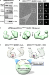
Fig. 4. DUB treatment of BRCA15382insC-BARD1 restores structural integrity.
(A) Western blot analysis of USP2-treated protein fractions isolated from HCC1937 cells experiencing oxidative stress. The band shift for BRCA15382insC to ~260 kDa in USP2-treated samples was confirmed by probing the BRCT and RING domains of BRCA1. Greater signal for the RING domain was detected in the USP2-treated samples along with a reduced signal for ubiquitin attachments at ~260 kDa. Increased levels of monoubiquitin (~8 kDa) were found in USP2-treated samples. (B) Image (left) and class averages (center) of mutated BRCA15382insC-BARD1 treated with 1 mM H2O2 and USP2. Scale bar, 50 nm. Projections of the 3D structure (right) agree with the class averages. Box size of averages, 25 nm. (C) The EM structure of BRCA15382insC-BARD1 shows a clamp-like motif lacking extra density adjacent to the RING domain (black circle) (fig. S4 and movie S4). Scale bar, 1.5 nm. (D) Difference peak (yellow) indicates the additional mass present in the hotspot region of BRCA15382insC under oxidative conditions. This area of extra mass is lacking in the USP2-treated BRCA15382insC-BARD1 structure (green).

References

    1. Welcsh P. L., Owens K. N., King M.-C., Insights into the functions of BRCA1 and BRCA2. Trends Genet. 16, 69–74 (2000). - PubMed
    1. Rowell S., Newman B., Boyd J., King M. C., Inherited predisposition to breast and ovarian cancer. Am. J. Hum. Genet. 55, 861–865 (1994). - PMC - PubMed
    1. Rosen E. M., Fan S., Ma Y., BRCA1 regulation of transcription. Cancer Lett. 236, 175–185 (2006). - PubMed
    1. Toft D. J., Cryns V. L., Minireview: Basal-like breast cancer: From molecular profiles to targeted therapies. Mol. Endocrinol. 25, 199–211 (2011). - PMC - PubMed
    1. Perou C. M., Molecular stratification of triple-negative breast cancers. Oncologist 15 (suppl. 5), 39–48 (2010). - PubMed

Publication types

MeSH terms

LinkOut - more resources