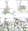
Potyvirus virion structure shows conserved protein fold and RNA binding site in ssRNA viruses
- PMID: 28948231
- PMCID: PMC5606705
- DOI: 10.1126/sciadv.aao2182
Potyvirus virion structure shows conserved protein fold and RNA binding site in ssRNA viruses
Abstract
Potyviruses constitute the second largest genus of plant viruses and cause important economic losses in a large variety of crops; however, the atomic structure of their particles remains unknown. Infective potyvirus virions are long flexuous filaments where coat protein (CP) subunits assemble in helical mode bound to a monopartite positive-sense single-stranded RNA [(+)ssRNA] genome. We present the cryo-electron microscopy (cryoEM) structure of the potyvirus watermelon mosaic virus at a resolution of 4.0 Å. The atomic model shows a conserved fold for the CPs of flexible filamentous plant viruses, including a universally conserved RNA binding pocket, which is a potential target for antiviral compounds. This conserved fold of the CP is widely distributed in eukaryotic viruses and is also shared by nucleoproteins of enveloped viruses with segmented (-)ssRNA (negative-sense ssRNA) genomes, including influenza viruses.
Figures


Similar articles
-
Structural basis for the multitasking nature of the potato virus Y coat protein.Sci Adv. 2019 Jul 17;5(7):eaaw3808. doi: 10.1126/sciadv.aaw3808. eCollection 2019 Jul. Sci Adv. 2019. PMID: 31328164 Free PMC article.
-
Structure of Turnip mosaic virus and its viral-like particles.Sci Rep. 2019 Oct 28;9(1):15396. doi: 10.1038/s41598-019-51823-4. Sci Rep. 2019. PMID: 31659175 Free PMC article.
-
The near-atomic cryoEM structure of a flexible filamentous plant virus shows homology of its coat protein with nucleoproteins of animal viruses.Elife. 2015 Dec 16;4:e11795. doi: 10.7554/eLife.11795. Elife. 2015. PMID: 26673077 Free PMC article.
-
Potyviral coat protein and genomic RNA: A striking partnership leading virion assembly and more.Adv Virus Res. 2020;108:165-211. doi: 10.1016/bs.aivir.2020.09.001. Epub 2020 Sep 18. Adv Virus Res. 2020. PMID: 33837716 Review.
-
Structural Homology Between Nucleoproteins of ssRNA Viruses.Subcell Biochem. 2018;88:129-145. doi: 10.1007/978-981-10-8456-0_6. Subcell Biochem. 2018. PMID: 29900495 Review.
Cited by
-
Structural basis for the multitasking nature of the potato virus Y coat protein.Sci Adv. 2019 Jul 17;5(7):eaaw3808. doi: 10.1126/sciadv.aaw3808. eCollection 2019 Jul. Sci Adv. 2019. PMID: 31328164 Free PMC article.
-
Turnip Mosaic Virus Coat Protein Deletion Mutants Allow Defining Dispensable Protein Domains for 'in Planta' eVLP Formation.Viruses. 2020 Jun 19;12(6):661. doi: 10.3390/v12060661. Viruses. 2020. PMID: 32575409 Free PMC article.
-
Structure of Turnip mosaic virus and its viral-like particles.Sci Rep. 2019 Oct 28;9(1):15396. doi: 10.1038/s41598-019-51823-4. Sci Rep. 2019. PMID: 31659175 Free PMC article.
-
Genome-Wide Variation in Potyviruses.Front Plant Sci. 2019 Nov 12;10:1439. doi: 10.3389/fpls.2019.01439. eCollection 2019. Front Plant Sci. 2019. PMID: 31798606 Free PMC article.
-
Genetic diversity analysis of papaya leaf distortion mosaic virus isolates infecting transgenic papaya "Huanong No. 1" in South China.Ecol Evol. 2020 Sep 22;10(20):11671-11683. doi: 10.1002/ece3.6800. eCollection 2020 Oct. Ecol Evol. 2020. PMID: 33144992 Free PMC article.
References
Publication types
MeSH terms
Substances
Supplementary concepts
LinkOut - more resources
Full Text Sources
Other Literature Sources
Miscellaneous

