. 2017 Nov 15;595(22):6923-6937.
doi: 10.1113/JP275142. Epub 2017 Oct 25.

The α2A -adrenoceptor suppresses excitatory synaptic transmission to both excitatory and inhibitory neurons in layer 4 barrel cortex

Affiliations

The α2A -adrenoceptor suppresses excitatory synaptic transmission to both excitatory and inhibitory neurons in layer 4 barrel cortex

Minoru Ohshima et al. J Physiol. .

Abstract

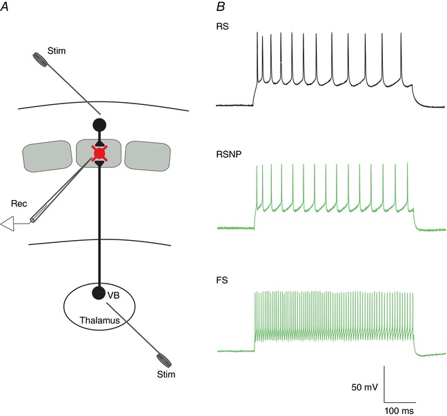
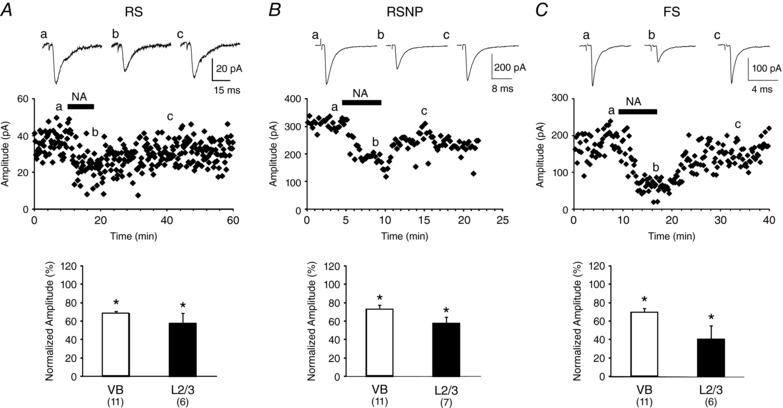
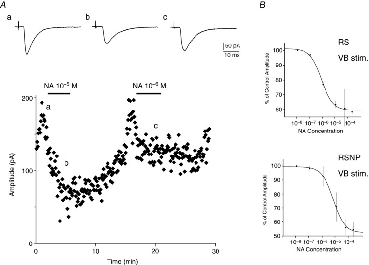
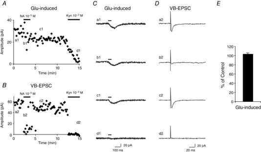
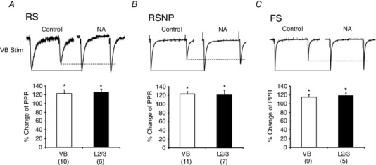
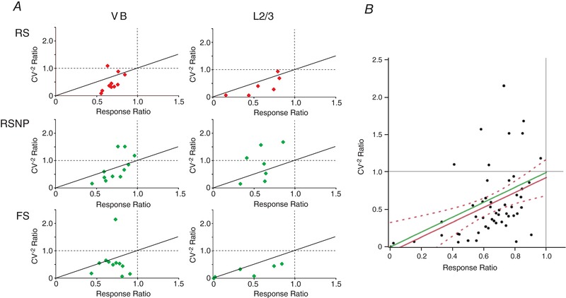
Key points: The effects of noradrenaline on excitatory synaptic transmission to regular spiking (excitatory) cells as well as regular spiking non-pyramidal and fast spiking (both inhibitory) cells in cortical layer 4 were studied in thalamocortical slice preparations, focusing on vertical input from thalamus and layer 2/3 in the mouse barrel cortex. Excitatory synaptic responses were suppressed by noradrenaline. However, currents induced by iontophoretically applied glutamate were not suppressed. Further, paired pulse ratio and coefficient of variation analysis indicated the site of action was presynaptic. Pharmacological studies indicated that the suppression was mediated by the α2- adrenoceptor. Consistent with this, involvement of α2A -adrenoceptor activation in the synaptic suppression in excitatory and inhibitory cells was confirmed by the use of α2A -adrenoceptor knockout mice.

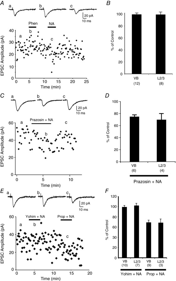
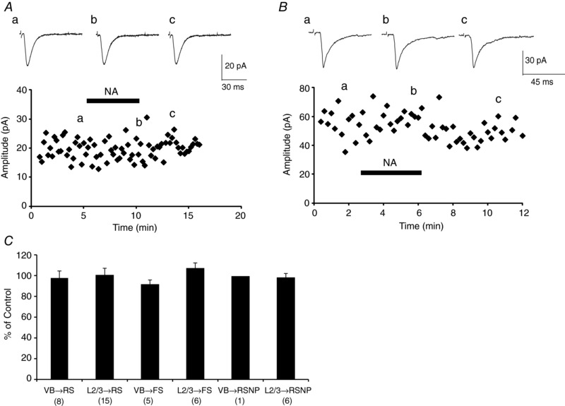
Abstract: The mammalian neocortex is widely innervated by noradrenergic (NA) fibres from the locus coeruleus. To determine the effects of NA on vertical synaptic inputs to layer 4 (L4) cells from the ventrobasal thalamus and layer 2/3 (L2/3), thalamocortical slices were prepared and whole-cell recordings were made from L4 cells. Excitatory synaptic responses were evoked by electrical stimulation of the thalamus or L2/3 immediately above. Recorded cells were identified as regular spiking, regular spiking non-pyramidal or fast spiking cells through their firing patterns in response to current injections. NA suppressed (∼50% of control) excitatory vertical inputs to all cell types in a dose-dependent manner. The presynaptic site of action of NA was suggested by three independent studies. First, responses caused by iontophoretically applied glutamate were not suppressed by NA. Second, the paired pulse ratio was increased during NA suppression. Finally, a coefficient of variation (CV) analysis was performed and the resultant diagonal alignment of the ratio of CV-2 plotted against the ratio of the amplitude of postsynaptic responses suggests a presynaptic mechanism for the suppression. Experiments with phenylephrine (an α1 -agonist), prazosin (an α1 -antagonist), yohimbine (an α2 -antagonist) and propranolol (a β-antagonist) indicated that suppression was mediated by the α2 -adrenoceptor. To determine whether the α2A -adrenoceptor subtype was involved, α2A -adrenoceptor knockout mice were used. NA failed to suppress EPSCs in all cell types, suggesting an involvement of the α2A -adrenoceptor. Altogether, we concluded that NA suppresses vertical excitatory synaptic connections in L4 excitatory and inhibitory cells through the presynaptic α2A -adrenoceptor.

Keywords: EPSC; alpha 2A-KO; mouse; noradrenaline; presynaptic inhibition; thalamocortical.


Figures

Figure 1
Figure 1. Experimental scheme and representative firing pattern of three different L4 cells
A, experimental scheme. Experiments were performed in thalamocortical slices. Recordings were made from excitatory and inhibitory cells in L4 barrels. Stimulation was applied to L2/3 or to the ventrobasal (VB) nucleus of the thalamus. Rec, recording electrode; Stim, stimulating electrode. B, three representative firing patterns of RS, RSNP and FS neurons in response to current injection.
Figure 2
Figure 2. Suppressive effect of NA on EPSC amplitude in three different L4 cell types
AC, top: examples of plots of EPSC amplitude during NA application (2 × 10−4 m) from an RS (A), RSNP (B) and FS (C) neuron in response to VB stimulation. Averaged traces (five trials) at the time indicated in the plots are shown above. Summaries of the effect of NA (2 × 10−4 m) on each cell type in response to VB and L2/3 stimulation are shown at the bottom.
Figure 3
Figure 3. Dose‐dependent suppression by NA
A, plot of EPSC amplitude recorded from an RS neuron by VB stimulation during NA application (10−5, 10−6 m). Averaged EPSCs (four trials) at the time indicated in the plot are shown above. B, dose–response curves of NA suppression of VB‐evoked EPSC from RS (top) and RSNP (bottom) neurons. EPSC amplitudes during NA administration were normalised by those of EPSC amplitude (mean ± SEM) before NA administration. Curve fit to a logistic equation by non‐linear regression.
Figure 4
Figure 4. Glutamate‐induced current was not suppressed by NA administration, indicating NA suppression is presynaptic
A, amplitudes of glutamate‐induced current were plotted against time. Administration of NA (10−5 m) barely suppressed glutamate‐induced current (b1). At the end of the experiment, kynurenate, a non‐selective glutamate receptor antagonist, was applied (1 mm), which successfully suppressed the glutamate‐induced current (d1), confirming that iontophoretically induced current was generated by direct activation of glutamate receptor. The clear difference between the effects of NA and kynurenate on glutamate‐induced current indicates that the site of action of NA is not postsynaptic. B, amplitudes of EPSCs evoked by VB stimulation applied 1500 ms after iontophoresis of glutamate (A). NA (10−5 m) suppressed VB‐evoked EPSCs (b2). Kynurenate (1 mm) also effectively eliminated VB‐evoked EPSCs (d2), as expected. C, trace examples of glutamate‐induced current at the time indicated in A. D, VB‐evoked EPSCs at the time indicated in B. E, quantification of glutamate‐induced current in the presence of NA (10−5 m). It became 103.5 ± 2.6% of control (n = 11, P > 0.1).
Figure 5
Figure 5. NA increased paired pulse ratio (PPR), suggesting that NA reduced presynaptic transmitter release
AC, recordings showing examples of PPR increases in RS (A), RSNP (B) and FS (C) cells are presented at the top. Paired pulses (100 ms intervals) were applied. EPSC amplitudes during NA administration (2 × 10−4 m) were scaled so that the first pulses of the control and NA administration matched. Summaries of PPR changes are shown at the bottom for EPSCs evoked by both VB and L2/3 stimulations.
Figure 6
Figure 6. Plots of the ratio of experimental to control CV−2 (where CV is coefficient of variation) as a function of the response ratio (EPSC amplitude in NA/EPSC amplitude in control) at an NA concentration of 2 × 10−4 m
A, results from individual cell types in response to VB or L2/3 stimulation. Continuous lines: results expected for a directly proportional response, indicating a presynaptic mechanism. B, pooled data from the different cell types and both VB and L2/3 stimulation. This plot fits the linear regression line: CV−2 = −0.067 + 0.998 × Response Ratio, which is indicated by a red line. This is almost aligned with the diagonal line (green; same as continuous lines in A), indicating a presynaptic mechanism. Dotted lines indicate 95% confidence intervals. The green diagonal line falls within this range, strongly supporting that the action is presynaptic.
Figure 7
Figure 7. Pharmacological studies indicating that α2‐ARs, but not α1‐ or β‐ARs are involved in the NA suppression
A, phenylephrine (10−4 m), an α1‐AR agonist, failed to suppress EPSCs evoked by VB stimulation in an RSNP cell, while subsequent NA application (10−5 m) suppressed the EPSCs in the same cell. Averaged traces (four trials) at the indicated time in the plots are shown above (a–c). B, summary of the effect of phenylephrine (10−4 m) in EPSCs evoked by VB and L2/3 stimulation. C, prazosin, an α1‐AR antagonist, at the concentration of 10−5 m failed to block suppression by NA that was simultaneously administered (10−5 m). Averaged traces (four trials) at the indicated time in the plots are shown above (a–c). D, summary of the effect of prazosin (10−5 m) in EPSCs evoked by VB and L2/3 stimulation. E, yohimbine, an α2‐AR antagonist, at the concentration of 5 × 10−5 m blocked NA suppression (10−5 m), while propranolol, a β‐AR antagonist (10−5 m), failed to block NA suppression (10−5 m). Averaged traces (four trials) at the indicated time in the plots are shown above (a–c). F, summary of the effect of yohimbine (5 × 10−5 m) and propranolol (10−5 m) in EPSCs evoked by VB and L2/3 stimulation.
Figure 8
Figure 8. NA (2 × 10−4 m) failed to suppress EPSCs in cells from α2A‐AR−/− mice
A and B, plots of EPSC amplitudes from an RS (A) and an FS (B) cell. Averaged traces (five trials) at the indicated time in the plots are shown at the top. C, summary of the effect of NA on all cell types in response to VB and L2/3 stimulations in cells from α2A‐AR−/− mice.

Similar articles

Cited by

References

    1. Agmon A & Connors BW (1991). Thalamocortical responses of mouse somatosensory (barrel) cortex in vitro. Neuroscience 41, 365–379. - PubMed
    1. Amaral DG & Sinnamon HM (1977). The locus coeruleus: Neurobiology of a central noradrenergic nucleus. Prog Neurobiol 9, 147–196. - PubMed
    1. Aoki C, Go CG, Venkatesan C & Kurose H (1994). Perikaryal and synaptic localization of α2A‐adrenergic receptor‐like immunoreactivity. Brain Res 650, 181–204. - PubMed
    1. Aston‐Jones G & Cohen JD (2005). An integrative theory of locus coeruleus‐norepinephrine function: Adaptive gain and optical performance. Annu Rev Neurosci 28, 403–450. - PubMed
    1. Bekkers JM & Stevens CF (1990). Presynaptic mechanism for long‐term potentiation in the hippocampus. Nature 346, 724–729. - PubMed

Publication types

MeSH terms

LinkOut - more resources