

Assessment of cerebrovascular development and intraventricular hemorrhages in preterm infants with optical measures of the brain arterial pulse wave
- PMID: 28949275
- PMCID: PMC6421243
- DOI: 10.1177/0271678X17732694
Assessment of cerebrovascular development and intraventricular hemorrhages in preterm infants with optical measures of the brain arterial pulse wave
Abstract
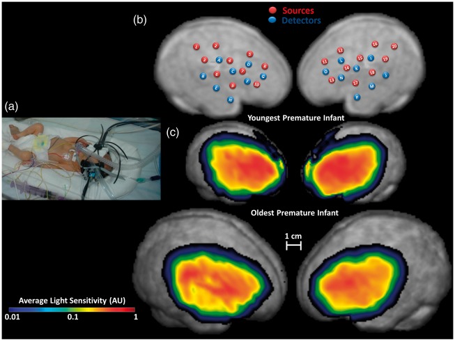
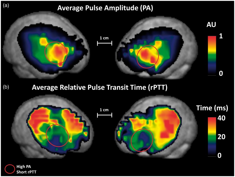
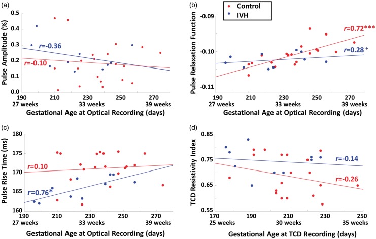
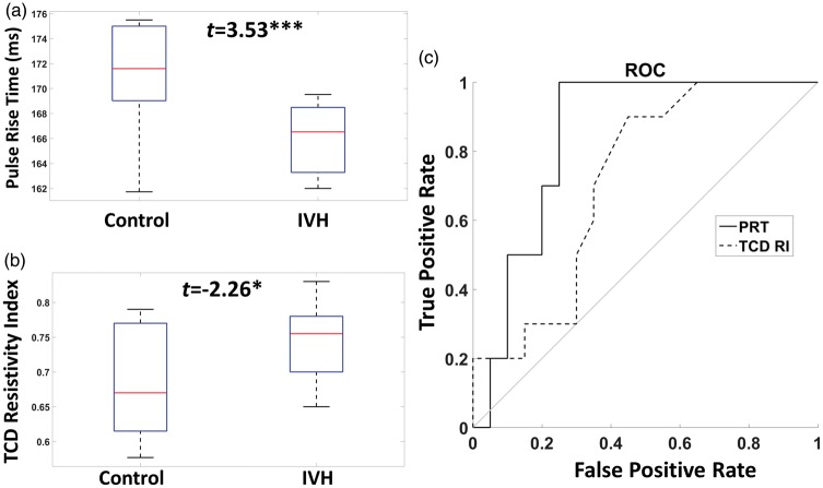
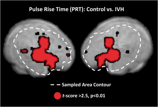
Preterm infants (born at 24-34 weeks of gestational age) suffer from a high incidence of neurological complications. Cerebrovascular lesions (intraventricular hemorrhages, IVH, and ischemic injury) due to the immaturity of the vascular system and its inability to adapt to the extra-uterine environment are the major causes of adverse neurological outcomes. We investigated the feasibility of assessing cerebrovascular status in preterm infants using a novel non-invasive optical procedure, pulse-DOT, usable within the incubator. Pulse-DOT, validated in adults, provides estimates of cerebral arterial status based on optical measurements of the pulse wave. These measurements are taken with a high-density optode montage and provide accurate spatial and temporal information. We found that two pulse parameters (pulse relaxation function, PReFx, and pulse rise time, PRT) in the investigated frontotemporal region, correlated with infant's age at recording, indexing cerebrovascular development. Moreover, PRT differentiated infants with and without concurrent IVH (sensitivity = 100%, specificity = 70%). These values are at least as high as those of the resistivity index obtained with transcranial Doppler of the middle cerebral artery, the current clinical method of choice for investigating arterial elasticity in preterm infants. This makes pulse-DOT a promising tool for investigating cerebrovascular risk factors and related pathologies in preterm infants.
Keywords: Preterm infants; brain arterial pulse wave (pulse-DOT); cerebrovascular development; diffuse optical tomography; intraventricular brain hemorrhages.
Figures

Similar articles
-
Cerebral oxygenation in preterm infants with germinal matrix-intraventricular hemorrhages.Stroke. 2010 Dec;41(12):2901-7. doi: 10.1161/STROKEAHA.110.597229. Epub 2010 Oct 21. Stroke. 2010. PMID: 20966409
-
Low superior vena cava flow and intraventricular haemorrhage in preterm infants.Arch Dis Child Fetal Neonatal Ed. 2000 May;82(3):F188-94. doi: 10.1136/fn.82.3.f188. Arch Dis Child Fetal Neonatal Ed. 2000. PMID: 10794784 Free PMC article.
-
Risk Factors for Intraventricular Hemorrhage in Preterm Infants Born at 34 Weeks of Gestation or Less Following Preterm Premature Rupture of Membranes.J Stroke Cerebrovasc Dis. 2016 Apr;25(4):807-12. doi: 10.1016/j.jstrokecerebrovasdis.2015.12.011. Epub 2016 Jan 18. J Stroke Cerebrovasc Dis. 2016. PMID: 26796051
-
Measuring cerebrovascular autoregulation in preterm infants using near-infrared spectroscopy: an overview of the literature.Expert Rev Neurother. 2017 Aug;17(8):801-818. doi: 10.1080/14737175.2017.1346472. Epub 2017 Jun 29. Expert Rev Neurother. 2017. PMID: 28639837 Review.
-
Cerebral vascular regulation and brain injury in preterm infants.Am J Physiol Regul Integr Comp Physiol. 2014 Jun 1;306(11):R773-86. doi: 10.1152/ajpregu.00487.2013. Epub 2014 Mar 19. Am J Physiol Regul Integr Comp Physiol. 2014. PMID: 24647591 Review.
Cited by
-
The Optical Effective Attenuation Coefficient as an Informative Measure of Brain Health in Aging.Photonics. 2019 Sep;6(3):79. doi: 10.3390/photonics6030079. Epub 2019 Jul 12. Photonics. 2019. PMID: 32377515 Free PMC article.
-
Optical measures of cerebral arterial stiffness are associated with white matter signal abnormalities and cognitive performance in normal aging.Neurobiol Aging. 2019 Dec;84:200-207. doi: 10.1016/j.neurobiolaging.2019.08.004. Epub 2019 Aug 10. Neurobiol Aging. 2019. PMID: 31500910 Free PMC article.
-
High-density diffuse optical tomography for imaging human brain function.Rev Sci Instrum. 2019 May;90(5):051101. doi: 10.1063/1.5086809. Rev Sci Instrum. 2019. PMID: 31153254 Free PMC article.
-
Noninvasive optical monitoring of cerebral hemodynamics in a preclinical model of neonatal intraventricular hemorrhage.Front Pediatr. 2025 Mar 10;13:1512613. doi: 10.3389/fped.2025.1512613. eCollection 2025. Front Pediatr. 2025. PMID: 40129699 Free PMC article.
-
Neonatal heart rate variability: a contemporary scoping review of analysis methods and clinical applications.BMJ Open. 2021 Dec 21;11(12):e055209. doi: 10.1136/bmjopen-2021-055209. BMJ Open. 2021. PMID: 34933863 Free PMC article.
References
-
- Martin JA, Kung H-C, Mathews TJ, et al. Annual summary of vital statistics: 2006. Pediatrics 2008; 121: 788–801. - PubMed
-
- Brew N, Walker D, Wong FY. Cerebral vascular regulation and brain injury in preterm infants. Am J Physiol Regul Integr Comp Physiol 2014; 306: R773–R786. - PubMed
-
- Doyle LW, Roberts G, Anderson PJ. Victorian Infant Collaborative Study Group. Outcomes at age 2 years of infants <28 weeks’ gestational age born in Victoria in 2005. J Pediatr 2010; 156: 49–53.e1. - PubMed
-
- Wolke D, Samara M, Bracewell M, et al. Specific language difficulties and school achievement in children born at 25 weeks of gestation or less. J Pediatr 2008; 152: 256–262. - PubMed
Publication types
MeSH terms
LinkOut - more resources
Full Text Sources
Other Literature Sources
Medical
