

Three-dimensional image reconstruction of distribution of Pnmt+ cell-derived cells in murine heart
- PMID: 28949324
- PMCID: PMC5613735
- DOI: 10.1038/sdata.2017.134
Three-dimensional image reconstruction of distribution of Pnmt+ cell-derived cells in murine heart
Abstract
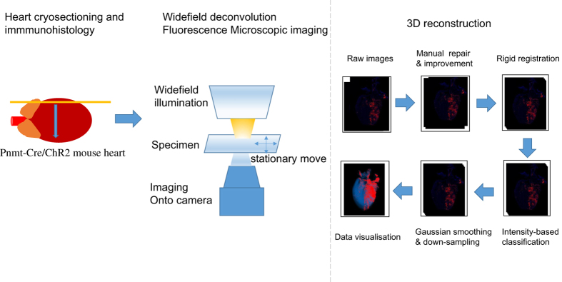
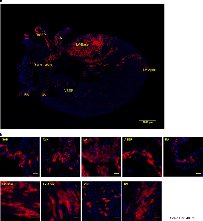
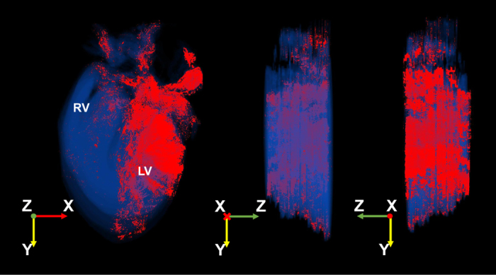
Elucidating the function of specific cell types in a highly complex multicellular system such as the heart often requires detailed anatomic reconstruction. We recently described a distinctive class of phenylethanolamine n-methyltransferase (Pnmt+) cell-derived cardiomyocytes (PdCMs), a new cardiomyocyte population with a potential endocrine role. In this dataset, a 3D reconstruction was carried out to visualise the distribution of PdCMs throughout the murine heart. Rigid registration (stiff rotation and translation) was applied to properly align the fused heart slice images based on landmarks using TrakEM2, an open source plug-in in Fiji. The registered slices were then analysed and reconstructed using MATLAB (MATLAB®. Version 8.3.0.532). The final reconstructed 3D volume was 561×866×48 pixels (corresponding to spatial resolutions of 5.8, 8.9 and 2.5 mm in the x-, y- and z-direction respectively), and visualised in Paraview. The reconstruction allows for detailed analyses of morphology, projections and cellular features of different cell types, enabling further geometrical and topological analyses. Image data can be accessed and viewed through Figshare.
Figures
Dataset use reported in
- doi: 10.1038/srep40687
Similar articles
-
Pnmt-Derived Cardiomyocytes: Anatomical Localization, Function and Future Perspectives.Front Physiol. 2019 Jul 10;10:713. doi: 10.3389/fphys.2019.00713. eCollection 2019. Front Physiol. 2019. PMID: 31354504 Free PMC article. Review.
-
Novel cardiac cell subpopulations: Pnmt-derived cardiomyocytes.Open Biol. 2020 Aug;10(8):200095. doi: 10.1098/rsob.200095. Epub 2020 Aug 19. Open Biol. 2020. PMID: 32810421 Free PMC article. Review.
-
High-Resolution 3D Heart Models of Cardiomyocyte Subpopulations in Cleared Murine Heart.Front Physiol. 2022 May 18;13:779514. doi: 10.3389/fphys.2022.779514. eCollection 2022. Front Physiol. 2022. PMID: 35665220 Free PMC article.
-
Optogenetic Control of Heart Rhythm by Selective Stimulation of Cardiomyocytes Derived from Pnmt+ Cells in Murine Heart.Sci Rep. 2017 Jan 13;7:40687. doi: 10.1038/srep40687. Sci Rep. 2017. PMID: 28084430 Free PMC article.
-
Distinctive left-sided distribution of adrenergic-derived cells in the adult mouse heart.PLoS One. 2011;6(7):e22811. doi: 10.1371/journal.pone.0022811. Epub 2011 Jul 27. PLoS One. 2011. PMID: 21818395 Free PMC article.
Cited by
-
A Protocol for Dual Calcium-Voltage Optical Mapping in Murine Sinoatrial Preparation With Optogenetic Pacing.Front Physiol. 2019 Aug 6;10:954. doi: 10.3389/fphys.2019.00954. eCollection 2019. Front Physiol. 2019. PMID: 31456689 Free PMC article.
-
Pnmt-Derived Cardiomyocytes: Anatomical Localization, Function and Future Perspectives.Front Physiol. 2019 Jul 10;10:713. doi: 10.3389/fphys.2019.00713. eCollection 2019. Front Physiol. 2019. PMID: 31354504 Free PMC article. Review.
-
Novel cardiac cell subpopulations: Pnmt-derived cardiomyocytes.Open Biol. 2020 Aug;10(8):200095. doi: 10.1098/rsob.200095. Epub 2020 Aug 19. Open Biol. 2020. PMID: 32810421 Free PMC article. Review.
-
Dbh+ catecholaminergic cardiomyocytes contribute to the structure and function of the cardiac conduction system in murine heart.Nat Commun. 2023 Nov 28;14(1):7801. doi: 10.1038/s41467-023-42658-9. Nat Commun. 2023. PMID: 38016975 Free PMC article.
-
High-Resolution 3D Heart Models of Cardiomyocyte Subpopulations in Cleared Murine Heart.Front Physiol. 2022 May 18;13:779514. doi: 10.3389/fphys.2022.779514. eCollection 2022. Front Physiol. 2022. PMID: 35665220 Free PMC article.
References
Data Citations
-
- Ni H. 2017. Figshare. https://doi.org/10.6084/m9.figshare.c.3692131 - DOI
References
-
- Ebert S. N. et al. Targeted insertion of the Cre-recombinase gene at the phenylethanolamine n-methyltransferase locus: A new model for studying the developmental distribution of adrenergic cells. Dev. Dyn. 231, 849–858 (2004). - PubMed
Publication types
MeSH terms
Substances
Grants and funding
LinkOut - more resources
Full Text Sources
Other Literature Sources
Molecular Biology Databases
Research Materials
Miscellaneous
